

Revista Portuguesa de Estomatologia, Medicina Dentária e Cirurgia Maxilofacial
SPEMD - SPEMD - Revista Portuguesa de Estomatologia Medicina Dentária e Cirurgia Maxilofacial | 2026 | 67 (2) | 91-98
Original research
Effect of remineralization agents on surface roughness of polyacid-modified composite resin: an in vitro study
Efeito de remineralizantes na rugosidade superficial de resina composta modificada por poliácido: um estudo in vitro
a Zonguldak Bülent Ecevit University, Faculty of Dentistry, Department of Pediatric Dentistry, Zonguldak, Turkey
Ebru Hazar Bodrumlu - hazarebru@yahoo.com
Article Info
Rev Port Estomatol Med Dent Cir Maxilofac
Volume - 67
Issue - 2
Original research
Pages - 91-98
Go to Volume
Article History
Received on 17/02/2026
Accepted on 21/06/2026
Available Online on 30/06/2026
Keywords
Original Research
Effect of remineralization agents on surfasse roughness of polyacid-modified composite resin: an in vitro study
Efeito de remineralizantes na rugosidade superficial de resina composta modificada por poliácido: um estudo in vitro
Ebru Hazar Bodrumlu1,* 0000-0002-3474-5583
Demet Demir Yıldırım1 0009-0008-9245-6625
1 Zonguldak Bülent Ecevit University, Faculty of Dentistry, Department of Pediatric Dentistry, Zonguldak, Turkey
Article history:
Received 17 February 2026
Accepted 21 June 2026
Available online 10 July 2026
Abstract
Objectives: Effective preventive measures are crucial for controlling dental caries, one of the most prevalent health problems in children. Polyacid-modified composite resin (PMCR) is commonly used in pediatric dentistry due to its aesthetic characteristics and ability to release fluoride. Surface roughness directly impacts the clinical success of this material. The present study aimed to assess the effects of various remineralizing agents on the surfasse roughness of PMCR.
Methods: A total of 48 PMCR discs were prepared and allocated into four experimental groups based on the remineralization agent applied: amorphous calcium phosphate (ACP), tricalcium phosphate (TCP), casein phosphopeptide–amorphous calcium phosphate with fluoride (CPP-ACP+F), and sodium fluoride (NaF). The discs’ surface roughness was measured using a profilometer before and after the agents were applied. Data distribution was assessed using the Shapiro–Wilk test, and statistical comparisons were performed using Welch’s ANOVA and paired-samples t-tests.
Results: The surface roughness did not differ significantly between groups before the application of the remineralizing agents (p=0.074). However, it increased significantly following application in all groups. The mean increase in arithmetical mean roughness was 0.400 ± 0.282 μm in the CPP-ACP+F group, 0.693 ± 0.647 μm in the NaF group, 0.439 ± 0.372 μm in the ACP group, and 0.953 ± 0.492 μm in the TCP group. In pairwise comparisons, the TCP group showed a significantly greater increase in surface roughness than the CPP-ACP+F group.
Conclusions: The present study’s findings indicate that exposure to the tested remineralization agents was associated with increased surface roughness values on PMCR surfaces under this accelerated exposure protocol. Furthermore, the magnitude of this increase varied depending on the remineralization agent used. Given that surface smoothness is a critical determinant of biological compatibility and the long-term performance of restorations, the potential adverse effects of remineralization agents should be considered in clinical practice.
Keywords: Calcium phosphates, Cariostatic agents, Composite resins, Fluorides, topical, Sodium fluoride, Tooth remineralization
Resumo
Objetivos: A aplicação de medidas preventivas eficazes é essencial para controlar a cárie dentária, uma das doenças mais prevalentes na infância. As resinas compostas modificadas por poliácido (PMCR) são amplamente utilizadas em odontopediatria devido às suas características estéticas e capacidade de libertar flúor. A rugosidade superficial influencia diretamente o sucesso clínico destes materiais. O presente estudo teve como objetivo avaliar os efeitos de diferentes agentes remineralizantes na rugosidade superficial de PMCR.
Métodos: Foram preparados 48 discos de PMCR e distribuídos por quatro grupos experimentais, de acordo com o agente remineralizante aplicado: fosfato de cálcio amorfo (ACP), fosfato tricálcico (TCP), fosfopeptídeo de caseína-fosfato de cálcio amorfo com flúor (CPP-ACP+F) e fluoreto de sódio (NaF). A rugosidade superficial dos espécimes foi avaliada com um perfilómetro, antes e após a aplicação dos agentes. A distribuição dos dados foi analisada pelo teste de Shapiro-Wilk e as comparações estatísticas foram efetuadas através da ANOVA de Welch e do teste t para amostras emparelhadas.
Resultados: A rugosidade superficial não diferiu significativamente entre os grupos antes da aplicação dos agentes remineralizantes (p=0,074). No entanto, aumentou significativamente após a aplicação em todos os grupos. O aumento médio da rugosidade média aritmética foi de 0,400±0,282 μm no grupo tratado com CPP-ACP+F, 0,693±0,647 μm no de NaF, 0,439±0,371 μm no de ACP e 0,953±0,491 μm no de TCP. Nas comparações par a par, o aumento observado no grupo tratado com TCP foi significativamente superior ao verificado no de CPP-ACP+F.
Conclusões: Os resultados do presente estudo indicaram que a aplicação de agentes remineralizantes esteve associada a um aumento da rugosidade superficial das PMCR. Além disso, a magnitude deste aumento variou consoante o agente remineralizante utilizado. Considerando que a lisura superficial constitui um determinante crítico da compatibilidade biológica e do desempenho a longo prazo das restaurações, os potenciais efeitos adversos dos agentes remineralizantes deverão ser tidos em consideração na prática clínica.
Palavras-chave: Fosfatos de cálcio,Agentes cariostáticos, Resinas compostas,Fluoretos tópicos,Fluoreto de sódio, Remineralização dentária
Introduction
Dental caries is among the most prevalent health problems in children, making effective preventive measures essential for its control and prevention.1 In recent years, a better understanding of the pathogenesis of caries and the identification of factors that influence remineralization and demineralization have increased the importance of preventive approaches. The main goal of preventive practice is to prevent demineralization of dental hard tissues and, where it has already occurred, to promote remineralization and thereby avert cavitation. Once demineralization advances and cavitation develops, the affected tooth must be restored.2, 3
Polyacid-modified composite resin (PMCR), a restorative material widely used in pediatric dentistry, was introduced in the early 1990s and developed to combine the superior aesthetic and mechanical properties of composite resins with the fluoride-releasing advantage of glass ionomer cements.4 - 7 It is widely used for both anterior and posterior restorations in primary teeth because of this combination of aesthetic, mechanical, and fluoride-releasing properties.8 However, the clinical performance of such materials is closely related not only to their mechanical durability but also to their surface properties.9
Among these properties, surface roughness is a key determinant of several unfavorable clinical outcomes, including plaque accumulation, discoloration, and material wear.10, 11
In the long term, a rough surface can increase plaque retention, leading to secondary caries, discoloration of the restoration surface, and inflammation of the surrounding soft tissues. Therefore, having a smooth surface and maintaining it over time has been regarded as one of the main criteria for the long-term success of restorative materials.11, 12 Various topical fluoride agents have been shown to cause surface alterations in restorative materials. Studies investigating the effects of remineralizing agents on the surface properties of these materials have reported material-dependent micromorphological changes; in particular, agents containing hydrofluoric and phosphoric acids have been shown to induce more pronounced surface degradation and to increase the surfasse roughness of many materials.13 - 15
Although remineralizing agents have been reported to induce certain surface changes, the surface roughness changes that may occur on PMCR following exposure to remineralizing agents have not been sufficiently investigated. Considering this gap in the literature, the present study aimed to evaluate the effects of the following remineralizing agents on the surfasse roughness of PMCR: amorphous calcium phosphate (ACP), tricalcium phosphate (TCP), casein phosphopeptide–amorphous calcium phosphate with fluoride (CPP-ACP+F), and sodium fluoride (NaF). The null hypothesis of this study was that the applied remineralizing agents would have no significant effect on PMCR’s surface roughness.
Materials and Methods
Four different remineralizing agents were selected due to their distinct chemical compositions and mechanisms of action, enabling a comprehensive comparison of their potential effects on the surface roughness of PMCR. The selected agentes were: ACP (Relief ACP Oral Care Gel, Philips Oral Healthcare,
Andover, MA, USA), also containing potassium nitrate and sodium fluoride in a gel-based carrier system; TCP (Citrine Varnish, Dharma Research, Inc., Miami, FL, USA), also containing 5% sodium fluoride (22,600 ppm fluoride) in a varnish-based carrier system; CPP-ACP+F (MI Varnish™ with RECALDENT™, GC Corporation, Tokyo, Japan), also containing 5% sodium fluoride (22,600 ppm fluoride) in a varnish-based carrier system; NaF (VOCO Profluorid Varnish, VOCO GmbH, Cuxhaven, Germany), corresponding to 5% sodium fluoride (22,600 ppm fluoride) in a colophony-based varnish carrier system with xylitol.
These agents are clinically relevant in pediatric dentistry, where they are commonly used to prevent and manage enamel demineralization and early caries lesions.
The PMCR used was Dyract XP (Dentsply Sirona, Charlotte, NC, USA) in capsule form, weighing 0.25 grams, and containing BIS-GMA, urethane methacrylate, TEGDMA, and butylated hydroxytoluene (BHT). Standard disc specimens prepared from the PMCR were used for surface roughness analyses. The sample size was calculated using G*Power software (version 3.1.9.2; Heinrich-Heine-Universität Düsseldorf, Germany) for a repeated-measures Welch’s ANOVA (within–between interaction).
Assuming a medium effect size (f = 0.25), an alpha of 0.05, and a power of 80%, a total of 48 specimens was required, corresponding to 12 specimens per group.
A total of 48 discs were fabricated, each with 10 mm in diameter and 2-mm thick. During the preparation of the PMCR discs, silicone molds with non-stick properties were used to ensure uniform and homogeneous shaping of the material.
After inserting the material into the mold, a transparent film was placed on the top surface of each disc, and a 100 mm × 100 mm × 4 mm glass plate was used to remove excess material, ensuring standard thickness and smoothness of the discs.
The polymerization of the disc specimens was performed using a portable LED light-curing device (Elipar S10, 3M ESPE, St. Paul, USA) for 20 seconds on each disc. Then, the surfaces of the discs were polished under dry conditions using the Opti-Disc polishing system (OptiDisc™, Kerr Corporation, Switzerland) with aluminum oxide discs in sequential particle sizes: 80 μm, 40 μm, 20 μm, and 10 μm. Each polishing disc was applied to the specimen surface for 10 seconds under light finger pressure. All polishing procedures were performed by the same operator, and a new disc was used for each specimen at each polishing step. Afterward, all samples underwent ultrasonic cleaning in distilled water for 10 minutes and were subsequently stored in a dry environment at 37 °C for 24 hours before baseline surface roughness measurements, to avoid variability associated with additional drying procedures and ensure standardized self-drying conditions for all specimens. The 48 PMCR discs prepared were equally allocated into four groups by an independent researcher using a simple randomization procedure. The allocation sequence was generated by the independent researcher—who was not involved in specimen preparation or surface roughness measurements—using a computer-generated list of random numbers. Group concealment was maintained by assigning a coded label to each specimen, so that the operator performing the pre- and post-application measurements remained blinded to group assignment. Following the allocation, each group was assigned a corresponding group label. The study groups were defined as follows: ACP group, TCP group, CPP-ACP+F group, and NaF group.
The baseline surface roughness of all specimens was measured using a TIME 3221 surface roughness tester (Beijing TIME High Technology Ltd., China). Prior to each measurement, the device was calibrated according to the manufacturer’s recommended calibration procedure. Measurements were performed using a 2-μm diamond stylus under an applied force of 0.75 mN. The cut-off length was set to 0.25 mm, the evaluation length to 1.25 mm, and the tracing speed to 0.25 mm/s.
At each of three predefined points on every disc specimen, a single tracing was recorded, and the mean of the three Ra (arithmetical mean roughness) values was taken as the pre-application surface roughness value. Each tracing was acquired over the 1.25-mm evaluation length, comprising five consecutive 0.25-mm sampling lengths. The Ra value for each point was automatically calculated as the average over these sampling lengths using the device’s standard Gaussian profile filter.
To ensure standardized measurement locations, one measurement point was selected at the center of each disc, while the other two points were positioned 2 mm from the center along the same axis in opposite directions. To maintain a consistente measurement orientation across all specimens, each specimen was positioned according to a predefined reference alignment, and the same trace orientation was used in both baseline and post-application measurements.
Based on a previously reported in vitro study simulating the clinical application protocols of intraoral remineralizing agents, each remineralizing agent was applied to the surfaces of the discs in the corresponding experimental group for four minutes.16 To simulate the cumulative effects of long-term, repeated clinical use rather than a single application, this procedure was repeated eight times for each specimen. These multiple cycles were deemed necessary to mimic prolonged exposure and detect potential surface alterations that may only become evident over time. According to the manufacturers’ recommendations, the remineralizing agents evaluated in this study are generally applied approximately two to four times per year in clinical practice. In the present study, assuming around four applications per year, eight application cycles were selected to represent approximately two years of clinical use. However, because the intervals normally occurring between clinical applications were not incorporated into the protocol, this approach should be considered an accelerated cumulative exposure model that may overestimate real-life exposure conditions. Each agent was applied according to the manufacturer’s instructions using a disposable applicator brush. The material was distributed over the entire exposed surface of each disc using gentle brushing strokes to obtain a thin, uniform layer.
Care was taken to ensure complete surface coverage while avoiding visible accumulation of the material. Following each application, the discs were rinsed with distilled water and dried, with no waiting period between applications.
After the treatment, the surface roughness of all discs was measured using the same method as before and recorded as the post-treatment measurements. All methodological procedures were carried out by a single operator to minimize operator-related variability.
Statistical analyses were performed using SPSS (version 26, IBM Corporation, New York, NY, USA) and Python (version 3.11.2; Python Software Foundation, Beaverton, OR, USA). The normality of the data was assessed using the Shapiro–Wilk test. Baseline surface roughness values were compared among the groups using Welch’s ANOVA because the assumption of homogeneity of variances was violated according to Levene’s test. Within-group differences between pre-application and post-application measurements were evaluated using paired-samples t-tests. The primary outcome measure was the change in surface roughness (ΔRa), calculated as the difference between post-application and pre-application Ra values. Since variance homogeneity was not satisfied, intergroup differences in ΔRa values were analyzed using Welch’s ANOVA followed by Tamhane’s T2 post-hoc test. The p-values obtained from within-group comparisons were considered descriptive and supportive, whereas the primary inferential focus of the study was based on between-group comparisons of ΔRa values. Statistical significance was set at p < 0.05.
Results
Analysis of the data revealed no statistically significant differences in the baseline surface roughness values among the groups (p = 0.074). This finding indicates that the pre-application surface characteristics were comparable.
In all examined groups, the surface roughness values measured after treatment differed significantly from those recorded before treatment (p < 0.05). The mean Ra value increased by 0.400 ± 0.282 μm (p < 0.001) in the CPP-ACP+F group, 0.693 ± 0.647 μm (p = 0.003) in the NaF group, 0.439 ± 0.372 μm (p = 0.002) in the ACP group, and 0.953 ± 0.492 μm (p < 0.001) in the TCP group. (Table 1) (Figure 1).
Table 1. Surface roughness measurements obtained at the pre-application and post-application stages of remineralizing agent application.
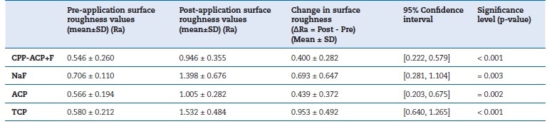
SD – standard deviation. Ra values are expressed in micrometers (μm).
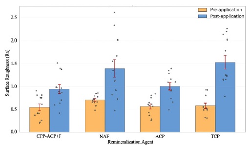
Figure 1. Comparison of mean surface roughness values (μm) of PMCR discs at pre-application and post-application stages (p < 0.05). Bars represent mean values, error bars indicate standard deviation (SD), and dots represente individual measurements
When pre- and post-application measurements were compared across groups, a statistically significant difference was observed only between the TCP and CPP-ACP+F groups (mean difference = 0.552 ± 0.164 μm; p = 0.021). The differences between the other group pairs were not statistically significant (p > 0.05) (Table 2).
Table 2. Tamhane’s post-hoc analysis for the statistical comparison of difference values between groups.
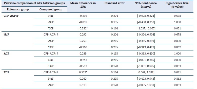
* Statistically significant difference between groups (p < 0.05).
Ra values are expressed in micrometers (μm).
Mean differences in ΔRa were calculated as the difference between the reference group and the compared group.
Discussion
Surface roughness, one of the mechanical surface properties of restorative materials, is defined as small surface irregularities that can negatively affect the long-term clinical success and performance of restorations, reduce their stability, increase their susceptibility to aging, and lead to aesthetic problems.17, 18 A review of the relevant literature shows that studies evaluating surface roughness have examined restorative materials with distinct mechanical and chemical properties, such as conventional glass ionomer cements, resin-modified glass ionomer cements, composite resins, and PMCR.17, 19 This study evaluated the effects of ACP, TCP, CPP-ACP+F, and NaF remineralizing agents on the surface roughness of PMCR, a material commonly used in pediatric dentistry.
This study’s results revealed that all remineralizing agents were associated with a statistically significant increase in PMCR’s surface roughness under this accelerated exposure protocol. Accordingly, the null hypothesis was rejected. In pairwise group analyses, TCP application resulted in a significantly greater increase in surface roughness than CPP-ACP+F. The increase did not differ significantly among the remaining groups. These findings are consistente with previous reports suggesting an association between exposure to fluoride- and calcium/phosphate-containing remineralizing agents and surface alterations in restorative materials.15, 19 - 21
Moreover, the greater increase in surface roughness observed in the TCP group compared to the CPP-ACP+F group may be related to differences in their mechanisms of action.
A previous study investigating surface alterations in demineralized enamel surfaces following the application of agentes containing β-TCP-F and CPP-ACP+F found that both reduced surface roughness.22 However, the β-TCP-F agent reduced surface roughness less than the CPP-ACP+F agent. The CPP-ACP+F agent was reported to stabilize calcium and phosphate ions and facilitate their delivery to the enamel surface, whereas, following application, β-TCP-F-containing varnishes release calcium, phosphate, and high concentrations of fluoride ions at the tooth surface as the varnish matrix is broken down by saliva.
In the literature, 0.2 μm has been reported as the critical surface roughness threshold for surfaces with no plaque accumulation; exceeding this value is associated with increased plaque retention and a consequent rise in the risk of caries and periodontal inflammation.9 Accordingly, a study conducted on implant abutments reported that an increase in surface roughness exceeding 0.2 μm significantly enhances plaque accumulation.23 In the present study, all remineralizing agents were associated with an increase in surface roughness exceeding 0.2 μm. Similarly, studies evaluating glass ionomer cements have reported surface roughness values predominantly exceeding 0.2 μm, whereas values below this threshold have generally been limited to surfaces produced by setting the material against a matrix strip, which represent the smoothest surfaces attainable and are unlikely to be reproduced under routine clinical conditions.9, 24 However, it should be noted that the commonly cited 0.2-μm plaque-retention threshold has largely been derived from heterogeneous studies involving different materials, substrates, and experimental conditions. Furthermore, as in the present study, only surface roughness was evaluated, whereas clinical outcomes such as secondary caries development or gingival inflammation were not assessed.
Therefore, any inferences based on this threshold should be interpreted with caution.
Besides its biological implications, increased surface roughness may adversely affect the long-term clinical performance of restorative materials by promoting surface degradation and compromising esthetic properties over time. Similar concerns have also been reported in the literature for other restorative materials, such as composite resins and glass ionomer cements, where surface roughness has been identified as an important factor influencing material longevity and clinical success.20, 21, 24, 25 Accordingly, taking measures to reduce surface roughness after application may be beneficial.26
In the literature, ACP-containing systems have been reported to support remineralization by continuously releasing calcium and phosphate ions in the oral environment and promote the formation of stable apatitic tooth mineral. However, it has also been shown that factors such as inadequate cohesion between the filler and resin matrix, increased water sorption, and polymerization shrinkage in ACP-containing composites may adversely affect their physicochemical and mechanical properties. Furthermore, the chemical structure and composition of the resin matrix have been reported to directly influence the degree of vinyl conversion (DVC), water sorption, and ion release. Accordingly, remineralizing agents containing ACP and casein phosphopeptide may interact with the structure of restorative materials, altering their surface properties.25 Moreover, studies evaluating the short-and long-term effects of fluoride-containing and non-fluoride immersion media on different types of composite resins have reported that surface roughness is material-dependent: microfilled composites exhibit higher roughness values, whereas nanofilled and microhybrid composites show lower values.27
A previous study reported that the sticky layer formed on the surface by a varnish containing 5% NaF increased the surface roughness of restorative materials.21 Another study examining the effects of various topical fluoride treatments on the surface roughness of restorative materials observed a statistically significant difference in roughness values between the group treated with APF gel and the control group.13 The researchers attributed this difference to the presence of hydrofluoric acid and phosphoric acid in the composition of the
APF gel, which may have influenced the surface roughness of the restorative materials. These findings indicate that remineralizing agents may influence the surface roughness of restorative materials. An experimental study investigating the effect of applying a 5% NaF-containing varnish to PMCR at different temperatures on surface roughness showed that it increased surface roughness; however, this increase was not statistically significant among the different temperature groups.19 According to previous findings, the CPP-ACP agente tested for enamel remineralization significantly reduced enamel surface roughness (Ra).28 This indicates that while such agents can reduce roughness on enamel surfaces by promoting remineralization, they may conversely increase surface roughness on restorative materials such as PMCR, as shown in the present study. This difference is thought to stem from the structural and chemical differences between the natural hydroxyapatite- containing composition of enamel and the synthetic composition of PMCR.
Although surface roughness-reducing effects of these remineralizing agents on enamel have been reported in the literature, the increase in surface roughness observed on the PMCR surface in the present study suggests that these agents may induce unfavorable interactions with restorative materials containing an organic matrix. Specifically, the interactions between these agents and the structural components may lead to deterioration of surface morphology and increased roughness.
Therefore, these agents should also be carefully evaluated on restorative material surfaces regarding restoration durability and long-term esthetic outcomes.
Increased surface roughness has been reported to promote bacterial colonization, leading to plaque accumulation, the development of secondary caries, gingival irritation, and aesthetic problems.24 In turn, the polishing protocol applied to the restoration surface has been reported to increase the resistance of restorations against remineralizing agents and play a significant role in maintaining surface integrity.26 Accordingly, applying a polishing procedure to restoration surfaces both before and after the use of remineralizing agents known to increase surface roughness in PMCR may help reduce potential undesirable effects on restoration surfaces. However, it is recommended that future studies also evaluate polishing procedures and investigate their effects directly to establish clearer evidence.
The present study evaluated surface roughness measurements before and after the application of the tested agents.
However, considering that factors such as specimen handling, repeated measurements, rinsing and drying procedures, storage conditions, and time may also influence surface roughness, the observed changes should not be solely attributed to the applied remineralizing agents. In this context, the absence of a control group undergoing the same experimental procedures without exposure to any remineralizing agent may be an important limitation of the study. Furthermore, the pH values of the materials, which might influence surface roughness, were not measured, which may constitute a limitation. Additionally, because this study was an in vitro investigation, the oral environment could not be fully simulated. In particular, the specimens were conditioned and stored under dry conditions at 37 °C rather than in water or artificial saliva. Although dry storage was selected to standardize self-drying conditions and to minimize variability among specimens, it differs from clinically relevant moist storage and does not reproduce the hydrated intraoral environment. Storage in water or artificial saliva could promote water sorption and hygroscopic changes within the resin-based matrix of PMCR, potentially modifying the surface response to the remineralizing agents; the direction and magnitude of this effect on the present surface roughness outcomes remain uncertain and warrant further investigation.
This should be considered an important limitation of the study. In future research, it is recommended to employ more comprehensive in vitro models that better mimic the oral environment, such as incorporating artificial saliva, thermal cycling, or pH cycling protocols, or conducting in vivo studies to overcome these limitations.
Conclusion
The present study found that exposure to all tested remineralizing agents was associated with a statistically significant increase in the surface roughness of the PMCR under this accelerated exposure protocol. Given that achieving and maintaining a smooth surface in restorative materials is a fundamental requirement for both biological compatibility and long-term restorative success, the potential effects of remineralizing agents on restorative material surfaces should be considered in clinical practice.
References
1. Hugoson A, Koch G, Göthberg C, Helkimo AN, Lundin SA, Norderyd O, et al. Oral health of individuals aged 3–80 years in Jönköping, Sweden during 30 years (1973–2003). II. Review of clinical and radiographic findings. Swed Dent J. 2005;29:139–55.
2. Cochrane N, Cai F, Huq N, Burrow M, Reynolds E. New approaches to enhanced remineralization of tooth enamel. J Dent Res. 2010;89:1187–97. 96 rev port estomatol med dent cir maxilofac . 2026;67(2):91-98
3. Meyer J, Cattani-Lorente M, Dupuis V. Compomers: between glass-ionomer cements and composites. Biomaterials. 1998;19:529–39.
4. Kanık Ö, Türkün LŞ. Restoratif cam iyonomer simanlarda güncel yaklaşımlar. Ege Univ Dishek Fak Derg. 2016;37:54–65.
5. Andersson-Wenckert IE, Folkesson UH, van Dijken JW. Durability of a polyacid-modified composite resin (compomer) in primary molars: a multicenter study. Acta Odontol Scand. 1997;55:255–60.
6. Saravanan SM, Srinivasan D, Eagappan AS, Priyal SD. Comparative assessment of compomers and ormocers as pit and fissure sealants in permanent molars among children aged 7–9 years. Int J Clin Pediatr Dent. 2024;17:742–7.
7. El Meligy OA, Othman HI, Abudawood SN, Al Tuwirqi AA, Alkeheli MF, Abdelrehim TR, et al. Histological evaluation of polyacid-modified composite resin and conventional
composite resin used for primary molars restoration. Dent J. 2024;12:343.
8. Kraemer N, Frankenberger R. Compomers in restorative therapy of children: a literature review. Int J Paediatr Dent. 2007;17:2–9.
9. Bollen CM, Lambrechts P, Quirynen M. Comparison of surface roughness of oral hard materials to the threshold surface roughness for bacterial plaque retention: a review of the literature. Dent Mater. 1997;13:258–69.
10. Turssi CP, Ferracane JL, Serra MC. Abrasive wear of resin composites as related to finishing and polishing procedures. Dent Mater. 2005;21:641–8.
11. Aykent F, Yondem I, Ozyesil AG, Gunal SK, Avunduk MC, Ozkan S. Effect of different finishing techniques for restorative materials on surface roughness and bacterial adhesion. J Prosthet Dent. 2010;103:221–7.
12. Özdal TPG, Duruk G. Effect of nutrients and medicines on surface of compomer. Cumhuriyet Dent J. 2019;22:211–7.
13. Çehreli ZC, Yazici R, García-Godoy F. Effect of 1.23 percent APF gel on fluoride-releasing restorative materials. ASDC J Dent Child. 2000;67:330–7.
14. Turssi CP, de Magalhães CS, Serra MC. Effect of fluoride gels on micromorphology of resin-modified glass-ionomer cements and polyacid-modified resin composites. Quintessence Int. 2001;32:571–7.
15. Avşar A, Tuloglu N. Effect of different topical fluoride applications on the surface roughness of a colored compomer. J Appl Oral Sci. 2010;18:171–7.
16. Mikami W, Koizumi H, Kodaira A, Hiraba H, Yoneyama T, Matsumura H. Gloss and surface roughness of prepolymerized composite materials designed for posterior CAD/CAM crown restorations corroded with acidulated phosphate fluoride application. Dent Mater J. 2022;41:60–7.
17. Oral F, Biçer AZY. Dental seramiklerde yüzey pürüzlülüğü. ADO Klin Bil Derg. 2022;11:103–7.
18. Türel V. Restoratif dental materyallerin yüzey mekanik özellikleri. Atatürk Univ Dis Hek Fak Derg. 2015;25:77–82.
19. Çevik N, Bodrumlu EH. Effects of fluoride varnish on roughness of polyacid-modified composite resins at diferente temperatures: an in vitro study. J Adv Oral Res. 2024;15:86–90.
20. Papagiannoulis L, Tzoutzas J, Eliades G. Effect of topical fluoride agents on the morphologic characteristics and composition of resin composite restorative materials. J Prosthet Dent. 1997;77:405–13.
21. Salama FS, Schulte KM, Iseman MF, Reinhardt JW. Effects of repeated fluoride varnish application on different restorative surfaces. J Contemp Dent Pract. 2006;7:54–61.
22. Barrera-Ortega CC, Rodil SE, Silva-Bermudez P, Delgado-Cardona A, Almaguer-Flores A, Prado-Prone G. Fluoride casein phosphopeptide and tri-calcium phosphate treatments for enamel remineralization: effects on surface properties and biofilm resistance. Dent J. 2025;13:246–52.
23. Quirynen M, Bollen CM, Papaioannou W, Van Eldere J, van Steenberghe D. The influence of titanium abutment surface roughness on plaque accumulation and gingivitis: shortterm observations. Int J Oral Maxillofac Implants. 1996;11:169–78.
24. Pacifici E, Bossù M, Giovannetti A, La Torre G, Guerra F, Polimeni A. Surface roughness of glass ionomer cements indicated for uncooperative patients according to surface protection treatment. Ann Stomatol (Roma). 2014;4(3-4):250–8.
25. O’Donnell JN, Schumacher GE, Antonucci JM, Skrtic D. Structure-composition-property relationships in polymeric amorphous calcium phosphate-based dental composites. Materials. 2009;2:1929–54.
26. Butler CJ, Masri R, Driscoll CF, Thompson GA, Runyan DA, Von Fraunhofer JA. Effect of fluoride and 10% carbamide peroxide on the surface roughness of low-fusing and ultra low-fusing porcelain. J Prosthet Dent. 2004;92:179–83.
27. De Oliveira ALBM, Giro EMA, Garcia PPNS, Campos JÁDB, Phark JH, Duarte S Jr. Roughness and morphology of composites: influence of type of material, fluoride solution, and time. Microsc Microanal. 2014;20:1365–72.
28. Arafa A. Synergetic remineralization effectiveness of calcium, phosphate and fluoride based systems in primary teeth. Pediatr Dent J. 2017;27:65–71.
Ebru Hazar Bodrumlu
E-mail address: hazarebru@yahoo.com
CRediT authorship contribution statement
Ebru Hazar Bodrumlu: Conceptualization, Data curation, Formal analysis, Investigation, Methodology, Resources, Supervision, Validation, Visualization, Writing – original draft, Writing – review & editing. Demet Demir Yıldırım: Conceptualization, Formal analysis, Investigation, Methodology, Resources, Validation, Writing – original draft, Writing – review & editing.
Ethical disclosures
Protection of human and animal subjects. The authors declare that no experiments were performed on humans or animals for this study.
Confidentiality of data. The authors declare that they have followed their work center protocols on access to patient data and for its publication.
Right to privacy and informed consent. The authors have obtained the written informed consent of the patients or subjects mentioned in the article. The corresponding author is in possession of this document.
Conflict of interest
The authors have no conflicts of interest to declare.
1646-2890/© 2026 Sociedade Portuguesa de Estomatologia e Medicina Dentária. Published by SPEMD.
This is an open access article under the CC BY-NC-ND license (http://creativecommons.org/licenses/by-nc-nd/4.0/).