

Revista Portuguesa de Estomatologia, Medicina Dentária e Cirurgia Maxilofacial
| 1
Original research
Awareness of forensic odontology and child abuse recognition among dentists
Consciência sobre odontologia legal e reconhecimento de maus-tratos infantis entre dentistas
a Department of Pediatric Dentistry, Faculty of Dentistry, Dokuz Eylul University, Izmir, Türkiye
b Private Pediatric Dentistry Practice, Istanbul, Türkiye
c Department of Pediatric Dentistry, Istanbul Health and Technology University, Istanbul, Türkiye
Aliye Tugçe Gürca - aliyetugce.gurcan@deu.edu.tr
Article Info
Rev Port Estomatol Med Dent Cir Maxilofac
Volume -
Issue -
Original research
Pages - 1
Go to Volume
Article History
Received on 12/03/2026
Accepted on 27/06/2026
Available Online on 30/06/2026
Keywords
Clinical Case Report
Management of discolored maxillary anterior teeth with/without pulp canal obliteration: a case series
Tratamento de dentes anteriores superiores descolorados com/sem obliteração do canal pulpar: uma série de casos
Sinem Buse Özvatan1 0009-0005-0954-9264
Deniz Yanik Nalbantoğlu2,* 0000-0001-5676-0293
1 Dr, Süleyman Demirel University, Faculty of Dentistry, Department of Endodontics, Isparta, Türkiye,
2 Associate Professor, Süleyman Demirel University, Faculty of Dentistry, Department of Endodontics, Isparta, Türkiye
Article history:
Received 3 April 2026
Accepted 15 June 2026
Available online 3 July 2026
Abstract
Discoloration of the anterior teeth with pulp canal obliteration (PCO) presents a significant clinical and aesthetic challenge, often impacting an individual’s quality of life. This case series describes the clinical application of the walking bleaching technique in discolored maxillary anterior teeth with PCO, and reports the observed clinical outcomes and patient-reported aesthetic perceptions. This case series comprised 7 patients (6 females, 1 male) aged 19 to 42 years. A total of 9 teeth were treated. The chief complaint of all patients was discoloration. Discolored maxillary anterior teeth were managed with walking bleaching. A cervical barrier of 2-mm glass ionomer cement was applied. 35% hydrogen peroxide was placed for 1 to 5 appointments, 3 days apart, followed by calcium hydroxide [Ca(OH)2] for one week. Then, composite restoration was performed. Follow-up periods of up to 14 months showed no symptoms. In all teeth, improved tooth appearance was clinically observed, and patients reported satisfaction with the aesthetic outcome. The technique was applied in complex clinical scenarios, including cases with total PCO confirmed by conebeam computed tomography, where conventional endodontic access was not feasible. No clinical or radiographic evidence of symptomatic inflammation or cervical resorption was observed during follow-up. Within the limitations of this case series, walking bleaching with 35% hydrogen peroxide is a minimally invasive treatment option for discolored maxillary anterior teeth, including in total PCO. While clinicians must adhere to regional safety regulations, including the placement of a cervical barrier and Ca(OH)2, this approach offers a conservative alternative to prosthetic rehabilitation.
Keywords: Aesthetics, dental, Dental pulp calcification,Hydrogen peroxide,Tooth bleaching,Tooth discoloration
Resumo
A descoloração dos dentes anteriores associada à obliteração do canal pulpar (OCP) representa um desafio clínico e estético significativo que afeta frequentemente a qualidade de vida dos indivíduos. Esta série de casos descreve a aplicação clínica da técnica de branqueamento ambulatório neste contexto e relata os resultados clínicos observados, bem como as perceções estéticas dos pacientes. Esta série de casos incluiu 7 pacientes (6 mulheres e 1 homem), com idades entre 19 e 42 anos. A principal queixa dos pacientes era descoloração dentária. No total, trataram-se 9 dentes anteriores superiores descolorados com a técnica de branqueamento ambulatório. Colocou-se uma barreira cervical de 2 mm de cimento de ionómero de vidro. Aplicou-se peróxido de hidrogénio a 35% durante 1 a 5 sessões, com intervalos de 3 dias. Posteriormente, manteve-se hidróxido de cálcio [Ca(OH)2] intracoronalmente durante uma semana, seguido de restauração com resina composta. O acompanhamento, de até 14 meses, não revelou sintomas clínicos. Todos os dentes apresentaram melhoria na aparência dentária, e os pacientes relataram satisfação com o resultado estético. A técnica foi aplicada em cenários clínicos complexos, incluindo casos de OCP total confirmada por tomografia computadorizada de feixe cónico em que o acesso endodôntico convencional não era viável. Não se observaram evidências clínicas ou radiográficas de inflamação sintomática ou reabsorção cervical durante o período de acompanhamento. Apesar das limitações desta série de casos, o branqueamento ambulatório com peróxido de hidrogénio a 35% pareceu proporcionar resultados estéticos satisfatórios em dentes anteriores superiores descolorados, incluindo casos de OCP total. Embora os clínicos devam seguir as regulamentações locais e os protocolos de segurança, incluindo a utilização de barreira cervical e hidróxido de cálcio intracoronal, esta abordagem pode representar uma alternativa conservadora às reabilitações protéticas.
Palavras-chave:Estética, odontologia,Calcificação da polpa dentária,Peróxido de hidrogénio,Branqueamento dentário,Descoloração dentária
Introduction
Tooth deformities and discolorations directly affect oral health-related quality of life (OHRQoL), both functionally and aesthetically.1 OHRQoL is defined as a state of oral health that enables an individual to speak, socialize, and eat without discomfort, pathology, or embarrassment. Aesthetics, a key componente of OHRQoL, is closely linked to self-confidence, and patients frequently seek dental care to achieve natural-looking teeth.2 Dental aesthetic perception is influenced by multiple factors, including tooth alignment, shape, and color, as well as social and psychological determinants, particularly among young individuals.1, 2
Internal discoloration refers to changes in the natural color and optical properties of the tooth structure caused by factors originating from the enamel and/or dentin.3 Local factors include caries, pulp necrosis, trauma, intrapulpal hemorrhage, pulp obliteration, irrigating solutions, intracanal medications, root canal filling materials, bioceramic materials, and temporary or permanent restorative materials, while systemic causes include medications, genetic disorders, and fluorosis.3, 4
Since the discoloration originates within the tooth, it is typically managed using intracoronal bleaching techniques, including the walking bleach technique, a minimally invasive endodontic procedure. The walking bleaching technique is a conservative, cost-effective intracoronal bleaching method that is widely regarded as reliable, offering advantages such as reduced chair time and ease of application compared with other techniques, including thermocatalytic and combined internal–external approaches.5 - 7 However, its success largely depends on accurate identification of the underlying etiological factors, precise diagnosis, and appropriate selection and execution of the procedure.
Various materials have been suggested for walking bleaching, including pyrozone, hydrogen peroxide, sodium perborate, and sodium peroxide.5 Hydrogen peroxide is the most commonly used bleaching agent, despite its cytotoxic and tissue-damaging effects. Although no clear consensus exists regarding the optimal concentration of hydrogen peroxide, lower concentrations have been suggested to reduce the risk of adverse effects.
Pulp canal obliteration (PCO), also known as calcific metamorphosis, is characterized by excessive hard tissue formation in response to trauma, with grayish or yellowish discoloration as its most prominent clinical finding.8 PCO presents a particular clinical challenge because aesthetic complaints may occur despite otherwise asymptomatic teeth without periapical pathology.
In these situations, clinicians must decide between monitoring, prophylactic endodontic treatment, or minimally invasive aesthetic management. Current evidence generally favors a conservative follow-up approach; however, when discoloration causes significant aesthetic concern, prophylactic root canal treatment followed by walking bleaching has traditionally been proposed.8, 9 This therapeutic dilemma raises na important clinical question: can aesthetic management of discolored teeth with PCO be achieved through a minimally invasive strategy that avoids unnecessary endodontic intervention?
Although walking bleaching is a well-established treatment for discolored non-vital teeth, evidence regarding its application in teeth with partial or total PCO remains limited, especially in cases where conventional canal access is not possible.
Therefore, the present case series aims to demonstrate the clinical management and outcomes of walking bleaching using 35% hydrogen peroxide on maxillary anterior teeth with different etiologies of discoloration, including CBCT-confirmed total PCO managed without canal negotiation.
Case Report
This case series included patients who presented with discolored maxillary anterior teeth, with or without PCO, for whom walking bleaching was selected as a conservative treatment option after clinical and radiographic assessment. According to institutional regulations, ethical approval was not required for this retrospective case series involving the reporting of routinely performed clinical procedures using anonymized patient data. Written informed consent was obtained from all patients.
Inclusion criteria were as follows: maxillary anterior teeth with discoloration requiring treatment; walking bleaching performed with the described protocol; available baseline clinical and radiographic records; and follow-up data. Exclusion criteria were cases with a lack of follow-up data, incomplete clinical/ radiographic documentation, systemic disease, and patients in whom walking bleaching was not completed according to the protocol.
Case identification was performed retrospectively. Patients who underwent the described walking bleaching protocol between March 2024 and September 2025 were screened for eligibility.
Consecutive eligible cases with available follow-up data were included. During this period, ten patients met the clinical inclusion criteria. Three patients were excluded because follow-up data were unavailable, preventing assessment of treatment outcomes. The remaining seven patients were included in the present case series.
Case-series flow: Ten patients were screened and met the clinical inclusion criteria. Three patients were excluded because follow-up data were unavailable. Seven patients with complete records and follow-up information were included and underwent outcome assessment.
The procedures were performed at the Department of Endodontics, Süleyman Demirel University Faculty of Dentistry, Turkey. All patients were referred from the Department of Oral and Maxillofacial Radiology. All clinical procedures were carried out by a single clinician (S.B.O., a general dentist with 4 years of experience in endodontic practice), ensuring procedural consistency throughout the case series. The follow-up examinations were conducted by an endodontist (D.Y.N., na endodontist with 8 years of clinical experience).
Tooth numbering throughout the manuscript follows the World Dental Federation (FDI) two-digit system.
Case 1
A 36-year-old, systemically healthy female patient was referred to the endodontics clinic with a chief complaint of discoloration in her right maxillary lateral incisor (tooth 12). Dental history demonstrated a previous endodontic treatment within the last 3 years. Clinical examination revealed that tooth 12 had a composite filling with no tenderness to percussion or palpation, and grayish discoloration was observed.
Periapical radiography revealed a previous root canal filling and a partial PCO at the apical half. No pathology was detected in the periapical tissues. Since the previous root canal treatment was radiographically adequate and no periapical pathology was present, a conservative walking bleaching approach was preferred. The patient was informed about the possible complications of walking bleaching and provided written and verbal informed consent.
A rubber dam was placed to prevent the agent from contactin the gingiva. The previous restoration was removed. The access cavity was cleaned of restorative materials, gutta-percha, and root canal sealer remnants up to 2 mm below the canal orifice. A 2 mm layer of conventional glass ionomer cement (GIC) (Ketac Cem, 3M ESPE, Germany) was placed at the canal orifice.
As a bleaching agent, 35% hydrogen peroxide (Opalescence Endo, Ultradent Products Inc., South Jordan, USA) was placed in the pulp chamber, covered with Teflon tape, and sealed temporarily with conventional GIC. After two appointments, 3 days apart, for the agent application, the desired color change was achieved.
The agent was removed from the cavity, followed by placement of calcium hydroxide [Ca(OH)2] for one week. In the following appointment, the temporary restoration and Ca(OH)2 were removed, and the pulp chamber was rinsed with saline solution. Definitive restoration was performed without any additional delay interval.
A selective-etch strategy was used in all cases with a two-step self-etch adhesive system (Clearfil SE Bond, Kuraray Noritake Dental Inc., Japan), followed by placement of a composite resin (Dynamic Plus, President Dental, Munich, Germany) (Figure 1).
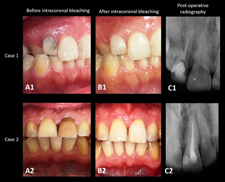
Figure 1. Clinical and radiographic sequence of walking bleaching in Case 1 (Tooth 12) and Case 2 (Tooth 21) with partial pulp canal obliteration. A1, A2 – Initial clinical appearance. B1, B2 – Postoperative aesthetic outcome. C1, C2 – Postoperative periapical radiograph.
Case 2
A 35-year-old, systemically healthy male patient was referred to the endodontic clinic with a chief complaint of discoloration in his left maxillary central incisor (tooth 21). Dental history revealed a previous root canal treatment 10 years earlier with no history of swelling or pain. Clinical examination showed a dark brown discoloration in tooth 21, with no percussion or palpation sensitivity. Periapical radiography showed no pathology. PCO was observed in the apical third of the root. Because the tooth was asymptomatic and the existing root canal treatment showed no radiographic deficiencies, only aesthetic management with walking bleaching was performed. The patient was informed about the possible complications of treatment and provided written informed consent.
The treatment procedure was performed as previously described. After 5 appointments performed 3 days apart, the desired color change was achieved. The agent was removed from the cavity, and Ca(OH)2 was applied for one week. Final restoration was performed using composite resin (Figure 1).
Case 3
A 36-year-old, systemically healthy female patient was referred to the endodontics clinic with a chief complaint of discoloration in her left maxillary central incisor (tooth 21). Clinical examination demonstrated a yellowish-light brown discoloration in tooth 21, and no other symptoms. Periapical radiography indicated healthy periapical tissues and total PCO.
Cone-beam computed tomography (CBCT) confirmed total PCO. Pulp sensitivity test demonstrated a negative response. Pulp necrosis was suspected based on the available clinical and radiographic findings. Considering the absence of symptoms and the CBCT-confirmed total PCO, aggressive canal negotiation was avoided to minimize the risk of iatrogenic complications, and a minimally invasive walking bleaching approach was selected. Accordingly, conventional root canal instrumentation and intracanal disinfection were not performed.
No attempt was made to negotiate the obliterated canal in order to avoid iatrogenic complications. Management was therefore limited to conservative access and walking bleaching.
A limited-access cavity was conservatively prepared within the obliterated pulp chamber area to create suficiente space for walking bleaching. Subsequently, a 2-mm GIC cervical barrier was placed at the cervical region, and 35% hydrogen peroxide was applied into the prepared coronal cavity above the cervical barrier without canal instrumentation. Walking bleaching procedures were performed as previously described. After two appointments performed 3 days apart, the desired color change was obtained, and the agent was removed. Ca(OH)2 was left in the cavity for one week, and a final composite resin restoration was placed (Figure 2).
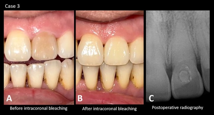
Figure 2. Management of Case 3 (Tooth 21) with total pulp canal obliteration. A – Initial clinical appearance. B – Clinical appearance after bleaching. C – Postoperative periapical radiograph
Case 4
A 40-year-old, systemically healthy woman was referred to the endodontic clinic with a chief complaint of discoloration in her maxillary central incisors (teeth 11 and 21). Dental history revealed a previous endodontic treatment 5 years earlier.
Clinical and radiographic examinations revealed yellow- brown discoloration without any additional symptoms. Radiographic evaluation of the periapical tissues and the previous root canal filling revealed no pathology or discrepancies.
As both teeth had acceptable previous endodontic treatment and no evidence of apical pathology, walking bleaching alone was considered sufficient to manage the aesthetic complaint for teeth 11 and 21. The patient was informed about the possible complications of this technique and provided informed consent.
The procedure was performed as previously described. After 4 appointments performed 3 days apart, the desired color change was achieved to the patient’s satisfaction. The agente was removed, and Ca(OH)2 was placed for one week, followed by placement of a composite resin restoration (Figure 3).
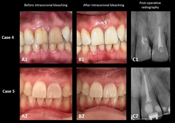
Figure 3. Treatment workflow of Case 4 (Teeth 11 and 21) and Case 5 (Tooth 21) without pulp canal obliteration. A1, A2 – Baseline clinical appearance. B1, B2 – Final aesthetic appearance after treatment. C1, C2 – Periapical radiography after treatment
Case 5
A 30-year-old, systemically healthy female patient presented to our clinic with a complaint of discoloration in her left maxillary central incisor (tooth 21). Clinical and radiological examination revealed dark hemorrhagic discoloration associated with pulp necrosis in tooth 21, and pulp sensitivity tests were negative. Root canal treatment was required because the tooth was non-vital and associated with discoloration secondary to pulp necrosis; walking bleaching was subsequently performed for aesthetic rehabilitation. The patient was informed about the possible complications of the treatment plan and provided written informed consent.
The access cavity was prepared under rubber dam isolation. The canal was instrumented using a rotary file system and irrigated using 2.5% sodium hypochlorite (NaOCl) and 17% ethylenediaminetetraacetic acid (EDTA) with ultrasonic irrigation activation (TA-200-S2H, Micron, Japan). Subsequently, the canal was filled using gutta-percha and epoxy resin sealer. A cervical barrier was placed using GIC. Walking bleaching was subsequently performed as previously described. After 3 days, the desired color was achieved. The agent was removed, and Ca(OH)2 was placed for one week.
Subsequently, the final restoration was made with composite resin (Figure 3).
Case 6
A 42-year-old, systemically healthy woman was referred to the endodontics clinic with a chief complaint of discoloration in her right maxillary lateral incisor (tooth 12). Clinical examination revealed gray discoloration in tooth 12. Pulp sensitivity tests (electric pulp test and cold test) were negative. Radiological examination showed mild changes in the periapical region.
Due to negative pulp sensitivity tests and mild periapical changes, root canal treatment was indicated before walking bleaching. The patient was informed about the possible complications of root canal treatment and walking bleaching and provided informed consent. Root canal treatment was performed as previously described.
Walking bleaching was performed for two appointments, 3 days apart. The desired color change was achieved, and the agent was removed from the cavity and dressed with Ca(OH)2 for 1 week. Then, the temporary restorations were removed, the pulp chamber was rinsed with saline, and the final restoration was made with composite resin (Figure 4).
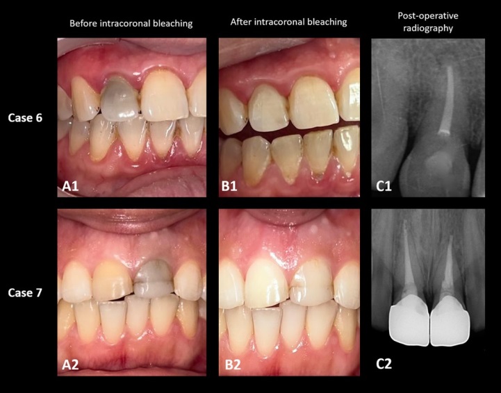
Figure 4. Aesthetic rehabilitation process of Case 6 (Tooth 12) and Case 7 (Teeth 11 and 21) without pulp canal obliteration. A1, A2 – Initial clinical presentation. B1, B2 – Postoperative clinical presentation. C1, C2 – Postoperative periapical radiography.
Case 7
A 19-year-old, systemically healthy woman was referred to the endodontic clinic with a complaint of discoloration in her left maxillary central incisor (tooth 21). The prosthodontic clinic planned a laminate veneer for teeth 11 and 21. Dental history revealed a previous root canal treatment 6 years earlier, with no history of pain or swelling. Clinical examination revealed gray discoloration in tooth 21. No tenderness to percussion or palpation was observed. Radiographic evaluation showed that teeth 11 and 21 had undergone root canal treatment, and no pathology was found. Thus, walking bleaching was performed as a conservative aesthetic procedure prior to planned laminate veneer restoration. The patient was informed about the possible complications of walking bleaching and provided informed consent.
After removal of the previous coronal restoration, gutta-percha remnants causing the discoloration in the pulp chamber were removed. A cervical barrier was placed. Walking bleaching was performed twice, 3 days apart, until the desired aesthetic was achieved. A satisfactory aesthetic outcome was achieved by the third appointment. The bleaching agent was removed from the cavity, and Ca(OH)2 was kept for 1 week.
Following completion of the walking bleaching procedure, a composite resin restoration was placed to provide coronal sealing and interim aesthetic rehabilitation before the planned laminate veneer treatment. The patient was subsequently referred to the prosthodontic clinic for definitive porcelain veneer restoration (Figure 4).
To standardize the walking bleaching protocol and minimize the risk of hydrogen peroxide diffusion-related complications, all cases were treated using the same clinical protocol.
In all teeth, gutta-percha and sealer remnants were removed up to 2 mm below the canal orifice when applicable, followed by placement of a 2-mm conventional GIC cervical barrier. The bleaching agent was restricted to the coronal cavity/pulp chamber above the cervical barrier and sealed with Teflon tape and temporary restoration material. Heat activation was not used in any case. Walking bleaching sessions were performed at standardized 3-day intervals, and calcium hydroxide dressing was maintained intracoronally for one week before definitive restoration.
To standardize cervical barrier positioning, gutta-percha was removed up to approximately 2 mm apical to the cemento- enamel junction (CEJ) using a bur with a known diameter.
Conventional GIC was then placed up to the canal orifice, creating approximately 2-mm-thick cervical barriers. In all cases, 35% hydrogen peroxide was confined within the pulp chamber/ coronal cavity without overflow and sealed temporarily with conventional GIC. The bleaching agent remained intracoronally during the 3-day inter-appointment period and was renewed at each session.
In this case series, the number of walking bleaching sessions ranged from 1 to 5 appointments, with most cases requiring 2–3 sessions to achieve clinically acceptable aesthetic improvement. In all cases, final color appearance was evaluated clinically by comparison with adjacent teeth under routine dental operatory lighting conditions.
An electric pulp tester (Pulp Tester; BeePower Taiwan Inc., Taiwan) was used to evaluate pulpal sensibility in all cases.
Periapical radiographs were acquired using a digital intraoral X-ray device (CS 2100, Carestream Dental LLC, Atlanta, GA, USA) set at 60 kV and 7 mA with an exposure time of 0.16 seconds. CBCT was acquired only in Case 3.
Follow-up
Seven patients were followed up for periods ranging from 6 to 14 months (Table 1). Follow-ups under 1 year were scheduled once, and follow-ups over 12 months were scheduled in 6-month periods. The predefined adverse outcomes assessed during follow-up included external cervical resorption, postoperative pain or sensitivity, development of periapical pathology, periodontal inflammation, restoration failure, and discoloration relapse.
Table 1. Summary of clinical characteristics, diagnosis, treatment approach, and follow-up outcomes of the presented cases.
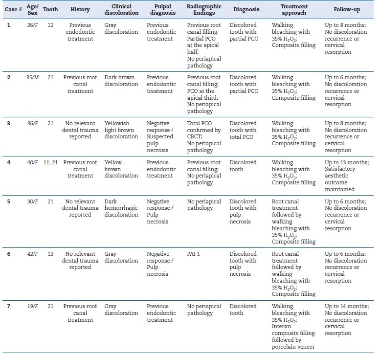
PAI – periapical index; PCO – pulp canal obliteration; H2O2 – hydrogen peroxide.
During follow-up, each case was examined clinically and radiographically. All periapical radiographs were taken according to the bisecting-angle periapical technique. No symptoms were observed in any case. At follow-up appointments, all treated teeth demonstrated stable aesthetic outcomes without clinically detectable discoloration recurrence. No gingival changes indicative of external cervical resorption (ECR) were observed.
Patients were satisfied with the aesthetic appearance, and no conditions requiring repeat treatment were observed. No predefined adverse events were observed in any of the cases. Moreover, periapical radiographs showed no radiographic findings suggestive of cervical resorption or periapical pathology. Demographic data, history, clinical and radiographic findings, treatment, and follow-up data for all cases are presented in Tables 1 and 2.
Table 2. Aesthetic outcomes and follow-up findings of the presented cases.
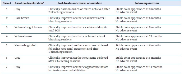
* Baseline discoloration and post-treatment appearance were assessed clinically and documented descriptively. No standardized shade guide, spectrophotometric assessment, or ΔE color analysis was performed.
PCO – pulp canal obliteration.
Discussion
The most important finding of this case series is that walking bleaching was not associated with clinically detectable adverse events during the reported follow-up period in teeth with partial or total PCO nor in endodontically treated teeth. The emphasis on aesthetic concerns, particularly in the anterior region, increases the clinical value of this treatment approach.
The literature shows that walking bleaching provides high aesthetic success and patient satisfaction, and that complication rates are low in case series.3, 5
Treatment planning in the present case series was primarily based on clinical symptoms, pulpal/periapical status, canal accessibility, and patient-centered aesthetic expectations.
Teeth presenting with previous adequate root canal treatment and no periapical pathology were managed conservatively with walking bleaching alone, whereas teeth with pulp necrosis or apical pathosis underwent root canal treatment before walking bleaching. In cases of PCO, particularly when total obliteration was confirmed by CBCT, avoidance of aggressive canal negotiation was preferred to minimize the risk of perforation and unnecessary loss of tooth structure. Therefore, treatment decisions were guided by a minimally invasive philosophy while maintaining aesthetic rehabilitation as the primary clinical objective.
In the present case series, all patients underwent a standardized diagnostic protocol including clinical examination and preoperative periapical radiographic assessment. Pulp sensibility tests were additionally performed in teeth without previous endodontic treatment, whereas previously treated teeth were evaluated according to the radiographic quality of the root canal filling, presence of symptoms, and periapical status. Non-surgical retreatment was not considered in asymptomatic teeth presenting with acceptable previous root canal treatment and partial PCO in the absence of periapical pathology. CBCT examination was selectively indicated in Case 3 because conventional radiographs were insufficient to determine the presence and trajectory of the canal system in a tooth with suspected total PCO. In contrast, CBCT was not considered necessary in cases of partial PCO because these teeth exhibited satisfactory root canal fillings, no radiographic peri-apical lesions, and no clinical symptoms other than aesthetic concerns. Therefore, additional three-dimensional imaging was not expected to alter treatment planning.
The management of PCO remains controversial, particularly in asymptomatic teeth with total canal calcification. A previous review reported that prophylactic endodontic treatment is generally not recommended in asymptomatic PCO cases without periapical pathology, supporting a conservative follow-up approach whenever possible.8 Furthermore, the authors emphasized that in cases of total PCO, invasive attempts to negotiate the canal may increase the risk of iatrogenic complications, and minimally invasive or external aesthetic management strategies should be preferred. In accordance with these recommendations, our approach prioritized the preservation of tooth structure and the avoidance of unnecessary endodontic intervention, especially in cases where total obliteration was confirmed by CBCT.
On the other hand, in the management of PCO cases, guided endodontic techniques have been proposed as minimally invasive alternatives for canal localization. Static and dynamic computer-aided approaches combine CBCT imaging with digital planning to improve access accuracy and reduce the risk of perforation or excessive dentin removal. Another previous review reported that guided endodontics may be particularly useful in anterior teeth with PCO.10 In the present case, canal negotiation was avoided due to total obliteration and the absence of symptoms. However, guided endodontic approaches may represent a valuable treatment option in selected cases requiring endodontic intervention.
Various bleaching agents are used for the walking bleaching technique.3, 5 These methods essentially involve the use of hydrogen peroxide-containing compounds in varying concentrations, formulations, and application systems. Among the most commonly used bleaching agents, hydrogen peroxide can be applied directly or produced by chemical reaction from sodium perborate or carbamide peroxide. Hydrogen peroxide releases free radicals, reactive oxygen species, and peroxide anions. Reactive molecules break down long-chain, dark-colored chromophore molecules and cleave double bonds into smaller, less colored, and more diffuse molecules.5, 6
The effects of various bleaching agents on the periodontal ligament and their potential to cause postoperative complications vary. Due to concerns about hydrogen peroxide use, some authors have advocated for the use of carbamide peroxide.7, 11
Carbamide peroxide, which is equivalent to 12% hydrogen peroxide when decomposed, has a low diffusion level and creates an alkaline environment by breaking down ammonia, making it a potentially safer alternative for walking bleaching.11 However, a recent study found similar effects of using 25% hydrogen peroxide and 37% carbamide peroxide in the walking bleaching technique on the levels of IL-1β and RANK-L markers in the gingival crevicular fluid profile over 12 months.12 The authors point out that clinicians should assess individual inflammatory tendencies when selecting a bleaching agent. In this case series, we closely evaluated patients’ periodontal status and susceptibility to inflammation during walking bleaching using 35% hydrogen peroxide. No periodontal inflammation was detected in any patient, nor were there any systemic diseases that could predispose to inflammation. According to our treatment results, the use of hydrogen peroxide in healthy periodontal tissues provided effective results and pallowed for timely definitive restoration.
ECR is one of the most critical complications associated with walking bleaching, particularly in teeth with a history of trauma and pre-existing hard tissue alterations. Although the exact mechanism is not fully elucidated, diffusion of hydrogen peroxide through dentinal tubules and CEJ defects is considered a key initiating factor, leading to inflammatory resorptive activity in susceptible cases.6 It has been suggested in the literature that higher concentrations of bleaching agents may increase the risk of this occurring, especially in the absence of an adequate cervical barrier.13 In the present case series, no clinical or radiographic evidence of cervical resorption was observed during the follow-up period, which may be attributed to the use of a properly sealed cervical barrier and controlled application protocol. However, given the limited follow-up duration, long-term monitoring remains essential to exclude late-onset resorptive changes. Furthermore, the absence of a standardized safety protocol may weaken conclusions regarding the safety of walking bleaching. Therefore, more stable clinical protocols are required to minimize the risk of ECR. On the other hand, it should be noted that ECR may be radiographically subtle in its early stages and may not always be detectable on conventional periapical radiographs; therefore, CBCT should be considered when clinical or radiographic findings raise suspicion of ECR.
Our results are similar to those of studies using 35% hydrogen peroxide. In a previous study, a combination of athome and in-office bleaching techniques was used in two cases; in both cases, patients did not report significant tooth sensitivity, and the treatment results met the patients’ expectations.14 In a case series of three patients, a 3–4 shade lightening was observed in vital upper central incisors after a single session of 35% hydrogen peroxide application, moderate to high levels of temporary tooth sensitivity were reported in all cases, and a 20–40% temporary decrease in pulp blood flow was observed with Laser Doppler, with both flow and sensitivity returning to baseline levels within one week.15 In a diferente study, 37% carbamide peroxide and 35% hydrogen peroxide were reported to have similar efficacy for non-vital walking bleaching.16
The success of walking bleaching treatment is multifactorial.
The severity of the initial discoloration, the type and concentration of the bleaching agent, the duration of application, the number of sessions, and the material used for the cervical barrier directly affect the treatment outcome.5 - 7 In our cases, repeated applications were required for teeth with intense discoloration, as in Case 2; however, satisfactory aesthetic results were obtained in all cases. According to our findings, light yellow and gray discolorations yielded results faster than dark yellow and black teeth.
The timing of restorative treatment after walking bleaching is also critical to treatment success. The presence of residual oxygen radicals in the pulp chamber may negatively affect the polymerization of composite resins and their bonding strength to dentin and enamel. It is recommended that restoration be performed 1-3 weeks later.17 In our cases, after the bleaching agent was removed, Ca(OH)2 was applied, and the restoration was postponed for one week. The most importante factor in permanent restoration after walking bleaching is preventing microleakage. Composite resins can prevent leakage by increasing the seal between the tooth and the restoration.
In our cases, this protocol was followed, and the final restorations were completed with composite resin. While walking bleaching is generally considered a safe procedure, its most important complication is ‘invasive cervical resorption’. Studies show that the thermocatalytic method using heat and the absence of a cervical barrier are associated with the occurrence of cervical resorption.6, 7, 18 Hydrogen peroxide placed in the pulp chamber spreads through the dentin tubules and enamel to reach the outer tooth surfaces and the periodontal tissues.19
Cervical resorption is mostly asymptomatic and is usually detected by routine radiographs. Clinically, it can sometimes manifest as swelling and percussion tenderness in the interdental papilla. Therefore, close follow-up is very important for treatment prognosis. A recent study reported significant increases in the levels of bone resorption markers RANK-L and IL-1β in the gingival crevicular fluid of teeth treated with 35% hydrogen peroxide or 37% carbamide peroxide as bleaching material, and these inflammatory mediators persisted for 3 months after walking bleaching.20, 21
Recommendations to reduce the risk of cervical-resorption complications include applying a cervical barrier at least 2 mm thick, avoiding the use of heat, limiting session durations, and preferring low-concentration agents.18 Various dental materials such as GICs, hydraulic filling materials (Cavit, Coltosol), resin composites, photo-activated temporary resin materials (Fermit), zinc oxide-eugenol cement, and zinc phosphate cement have been suggested as temporary restoration materials for cervical barriers.3, 6, 7 In our cases, cervical barrier application with conventional GIC was performed on all teeth, and no evidence of resorption was observed during the follow-up period.
In a previous study, a walking bleaching protocol combining 35% hydrogen peroxide and sodium perborate, along with the placement of a 2.5-mm cervical barrier, was applied to 255 teeth.22 During a follow-up period ranging from 6 months to 5 years, no cases of cervical resorption were reported. These findings suggest that the risk of cervical resorption may be more strongly associated with the presence of an adequate cervical barrier and the use of heat to potentiate hydrogen peroxide activity, rather than with the concentration of hydrogen peroxide alone.
Another preventive approach for cervical resorption is the placement of calcium hydroxide as an intracoronal medicamente following walking bleaching. Ca(OH)2 counteracts the acidic effects of hydrogen peroxide radicals by creating an alkaline environment, thereby reducing the risk of inflammatory root resorption.18 Maintaining Ca(OH)2 within the pulp chamber for one week after the walking bleaching procedure promotes alkalization of the surrounding tissues, which may help protect the periodontal ligament and prevent the development of cervical resorption.22 In all of our cases, definitive restorations were completed after a one-week Ca(OH)2 application period.
Regarding clinical contraindications, clinicians should follow local regulations and current clinical guidelines on the use of hydrogen peroxide-containing bleaching agents. Regulatory requirements governing the clinical use of high-concentration hydrogen peroxide vary across countries and jurisdictions, and restrictions may apply to the concentration, indication, or mode of application of bleaching products. Therefore, the clinical protocol described in the present case series should be interpreted within the regulatory framework applicable at the time and place of treatment, and its applicability may differ according to local legal requirements and professional recommendations.
The clinical procedures described in this case series were performed in accordance with the regulations and professional standards applicable at the time and place of treatment. In addition, walking bleaching procedures should be carefully planned for patients with potential contraindications, including pregnancy and young age.18, 22, 23
PCO presents a significant clinical challenge due to the extensive deposition of tertiary dentin within the pulp chamber and root canals. While the primary clinical manifestation of this entity is a yellowish-brown or grayish discoloration, the management of such cases often leads to a therapeutic dilemma.
Traditional protocols suggest proactive root canal treatment to facilitate walking bleaching; however, the absence of a discernible canal path increases the risk of iatrogenic complications, such as lateral perforations. In this case series, a minimally invasive strategy was employed, particularly in cases where total obliteration was confirmed via CBCT. CBCT is a valid diagnostic tool for determining root canal morphology and its relationship with surrounding tissues. In teeth with PCO, a comprehensive CBCT evaluation and determination of canal presence and trajectory before walking bleaching procedures are important for prognosis.
Interpretation of pulp sensibility tests in teeth with PCO requires caution. Sensibility tests assess neural response rather than true pulpal vitality, and false-negative responses may occur due to extensive calcification and reduced neural conductivity.
Therefore, negative sensibility test results should not be considered diagnostic of pulp necrosis in isolation and must be interpreted in conjunction with clinical findings, radiographic assessment, and the presence or absence of periapical pathosis.
Calcium silicate-based materials such as MTA and Biodentine have been associated with coronal tooth discoloration, especially in the presence of blood contamination. Previous studies reported that regenerative and vital pulp therapy procedures using these materials may result in clinically visible discoloration over time.4, 24 Therefore, discoloration observed in endodontically treated teeth may not only be related to pulp necrosis or restorative materials, but also to the use of calcium silicate-based cements. This should be considered during the differential diagnosis and treatment planning of walking bleaching cases.
One limitation of the present case series is the lack of objective shade assessment and standardized outcome measures. Aesthetic outcomes were evaluated using clinical examination and patient satisfaction, without standardized color-measurement methods, including VITA shade guides, spectrophotometers, or ΔE analysis. Therefore, the predictability of color change could not be quantitatively assessed. Future studies with standardized shade-evaluation protocols and larger sample sizes are needed to provide more objective data on bleaching outcomes. In addition, patient satisfaction was documented qualitatively during routine follow-up visits and was not assessed using a standardized patient-reported outcome measure such as a visual analog scale or Likert-scale questionnaire. Future studies should incorporate validated patient-reported outcome measures to provide a more systematic assessment of patient satisfaction and perceived aesthetic improvement. Furthermore, periapical radiographs were obtained using the bisecting-angle technique as part of routin clinical practice. Although this method is widely used clinically, it may provide less reproducible projection geometry than the paralleling technique during longitudinal follow-up assessments.
This should be considered when interpreting radiographic comparisons over time.
Other limitations included a small sample size and the absence of a control or comparator group, limiting the strength of causal inferences. Although all eligible cases with available follow-up data were included, three otherwise eligible patients were excluded because follow-up information was unavailable, which may have introduced selection bias. The small sample size and single-center nature of the case series may also limit representativeness and external validity. Moreover, the included cases exhibited heterogeneous clinical characteristics, which may have influenced treatment outcomes. In turn, the relatively short follow-up period limits the ability to detect rare but clinically relevant complications, such as ECR.
Finally, the generalizability of the findings is constrained, particularly due to regional regulations restricting hydrogen peroxide concentrations, which may differ from protocols reported in the broader literature.
Our findings suggest that even in the absence of traditional endodontic access, walking bleaching can be effectively performed provided that a secure cervical barrier is established to mitigate the risk of cervical resorption. No clinical or radiographic adverse events were observed during the reported follow-up period in this limited case series. Within the limitations of this descriptive case series, walking bleaching produced satisfactory aesthetic outcomes in the reported cases.
This approach aligns with the ‘watchful waiting’ philosophy, prioritizing the preservation of tooth structure while addressing the patient’s aesthetic expectations.
Conclusion
Within the limits of the short follow-up period in this case series, walking bleaching with 35% hydrogen peroxide was associated with satisfactory clinical outcomes in teeth with both partial and total PCO. In the case where total PCO was confirmed by CBCT and conventional canal access was not feasible, a CBCT-informed conservative approach was adopted.
However, due to the limited sample size and follow-up duration, these results should be interpreted cautiously. Furthermore, strict adherence to the clinical protocol is crucial, including placement of a 2-mm cervical barrier and a one-week intracoronal calcium hydroxide dressing. Given the lack of objective shade assessment, the small number of included cases, and the relatively short follow-up period, further studies with standardized outcome measures and longer-term follow- up are required to better characterize the clinical outcomes of this treatment approach.
References
1. Kovacevic Pavicic D, Pavlic A, Kinkela Devcic M, Lajnert V, Spalj S. Tooth color as a predictor of oral health-related quality of life in young adults. J Prosthodont. 2019;28:886-92.
2. Meireles SS, Goettems ML, Dantas RV, Bona ÁD, Santos IS, Demarco FF. Changes in oral health-related quality of life after dental bleaching in a double-blind randomized clinical trial. J Dent. 2014;42:114-21.
3. Bersezio C, Ledezma P, Torres V, Villagra J, Aravena D, Kuga M, Martín J, Fernandez E. Comparative efficacy of In-office and walking bleach techniques in whitening of non-vital teeth. Clin Oral Investig. 2024;29:24.
4. Ramos JC, Palma PJ, Nascimento R, Caramelo F, Messias A, Vinagre A, Santos JM. 1-year In vitro evaluation of tooth discoloration induced by 2 calcium silicate-based cements. J Endod. 2016;42:1403-7.
5. Das G, Qasim M, Rana MH, Sharif RA, Suleman G, Sainudeen S, Khader MA, Javali MA, Ishfaq M, Ibrahim M, Shah SJ, Khalid I. Comparative efficacy of external dental bleaching gels: a systematic review of whitening performance and longevity. Clin Oral Investig. 2025;29:546.
6. Newton R, Hayes J. The association of external cervical resorption with modern internal bleaching protocols: what is the current evidence? Br Dent J. 2020;228:333-7.
7. Lee GP, Lee MY, Lum SO, Poh RS, Lim KC. Extraradicular diffusion of hydrogen peroxide and pH changes associated with intracoronal bleaching of discoloured teeth using different bleaching agents. Int Endod J. 2004;37:500-6.
8. Vinagre A, Castanheira C, Messias A, Palma PJ, Ramos JC. Management of pulp canal obliteration—systematic review of case reports. Medicina (Kaunas). 2021;57:1237.
9. Bastos JV, Côrtes MIS. Pulp canal obliteration after traumatic injuries in permanent teeth—scientific fact or fiction? Braz Oral Res. 2018 Oct 18;32:75.
10. Ribeiro D, Reis E, Marques JA, Falacho RI, Palma PJ. Guided Endodontics: Static vs. Dynamic Computer-Aided techniques—a literature review. J Pers Med. 2022;12:1516.
11. Lim MY, Lum SO, Poh RS, Lee GP, Lim KC. An in vitro comparison of the bleaching efficacy of 35% carbamide peroxide with established intracoronal bleaching agents. Int Endod J. 2004;37:483-8.
12. Miranda CB, Mayer C, Ledezma P, Vernal R, Rivera O, Godoy EF. Persistent inflammatory response in periodontal tissues after walking bleach: a clinical biomarker-based study. Aust Endod J. 2025;51:619-31.
13. Heboyan A, Avetisyan A, Karobari MI, Marya A, Khurshid Z, Rokaya D, Zafar MS, Fernandes GVO. Tooth root resorption: a review. Sci Prog. 2022;105:368504221109217.
14. Ferretti MA, Kury M, Mendonça BCD, Giannini M, Cavalli V, Aguiar FHB. Combination of at-home and in-office bleaching techniques: case series. Rev Gaúch. Odontol. 2021;69:1-7.
15. Cartagena AF, Parreiras SO, Loguercio AD, Reis A, Campanha NH. In-office bleaching effects on the pulp flow and tooth sensitivity—case series. Braz Oral Res. 2015;29:1-6.
16. Knezevic N, Obradovic M, Dolic O, Veselinovic V, Kojic Z, Josipovic R, Arapovic-Savic M. Clinical testing of walking bleach, in-office, and combined bleaching of endodontically treated teeth. Medicina (Kaunas). 2022;59:1-13.
17. Cavalli V, Reis AF, Giannini M, Ambrosano GM. The effect of elapsed time following bleaching on enamel bond strength of resin composite. Oper Dent. 2001;26:597-602.
18. Attin T, Paqué F, Ajam F, Lennon AM. Review of the current status of tooth whitening with the walking bleach technique. Int Endod J. 2003;36:313-29.
19. Palo RM, Bonetti-Filho I, Valera MC, Camargo CH, Camargo S, Moura-Netto C, Pameijer C. Quantification of peroxide ion passage in dentin, enamel, and cementum after internal bleaching with hydrogen peroxide. Oper Dent. 2012;37:660-4.
20. Bersezio C, Vildósola P, Sáez M, Sánchez F, Vernal R, Oliveira OB Jr, Jorquera G, Basualdo J, Loguercio A, Fernández E. Does the use of a walking bleaching technique increase boné resorption markers? Oper Dent. 2018;43:250-60.
21. Bersezio C, Sánchez F, Estay J, Ledezma P, Vernal R, Garlet G. Inflammatory markers IL1β and RANK-L assessment after non-vital bleaching: a 3-month follow-up. J Esthet Restor Dent. 2020;32:119-26.
22. Machado AC, Braga SRM, Ferreira D, Jacintho FF, Scaramucci T, Sobral MAP. Bleaching of severely darkened nonvital tooth case report—48 months clinical control. J Esthet Restor Dent. 2021;33:314-22.
23. Abbott P, Heah SY. Internal bleaching of teeth: an analysis of 255 teeth. Aust Dent J. 2009;54:326-33.
24. Palma PJ, Marques JA, Falacho RI, Correia E, Vinagre A, Santos JM, Ramos JC. Six-month color stability assessment of two calcium silicate-based cements used in regenerative endodontic procedures. J Funct Biomater. 2019;10:14.
Deniz Yanık Nalbantoğlu
E-mail address: deniznalbantoglu@sdu.edu.tr
CRediT authorship contribution statement
Sinem Buse Özvatan: Conceptualization; Data curation; Formal analysis; Funding acquisition; Validation; Visualization; Writing – original draft. Deniz Yanık Nalbantoğlu: Data curation; Methodology; Supervision; Validation; Writing – original draft; Writing – review & editing.
Ethical disclosures
Protection of human and animal subjects. The authors declare that the procedures followed were in accordance with the regulations of the relevant clinical research ethics committee and with those of the Code of Ethics of the World Medical Association (Declaration of Helsinki).
Confidentiality of data. The authors declare that no patient data appear in this article.
Right to privacy and informed consent. The authors have obtained the written informed consent of the patients or subjects mentioned in the article. The corresponding author is in possession of this document.
Conflict of interest
The authors have no conflicts of interest to declare. Ethical disclosures
1646-2890/© 2026 Sociedade Portuguesa de Estomatologia e Medicina Dentária. Published by SPEMD.
This is an open access article under the CC BY-NC-ND license (http://creativecommons.org/licenses/by-nc-nd/4.0/).