
Revista Portuguesa de Estomatologia, Medicina Dentária e Cirurgia Maxilofacial
SPEMD - SPEMD - Revista Portuguesa de Estomatologia Medicina Dentária e Cirurgia Maxilofacial | 2026 | 67 (2) | 63-70
Original research
Ozone-activated platelet-rich plasma combined with occlusal splint therapy in the treatment of temporomandibular joint osteoarthritis
Plasma rico em plaquetas ativado por ozono combinado com férula oclusal na osteoartrite temporomandibular
a Manuel Ascunce Domenech University Hospital, Camagüey, Cuba
b University of Medical Sciences of Camagüey, Camagüey,Cuba.
Reynier Ramírez - reynierramirez93@gmail.com
Article Info
Rev Port Estomatol Med Dent Cir Maxilofac
Volume - 67
Issue - 2
Original research
Pages - 63-70
Go to Volume
Article History
Received on 22/03/2026
Accepted on 04/06/2026
Available Online on 30/07/2026
Keywords
Original Research
Ozone-activated platelet-rich plasma combined with occlusal splint therapy in the treatment of temporomandibular joint osteoarthritis
Plasma rico em plaquetas ativado por ozono combinado com férula oclusal na osteoartrite temporomandibular
Reynier Ramírez1,2,* 0000-0002-3759-0249
Yoan R. Arango1 0009-0005-3742-8443
1 Manuel Ascunce Domenech University Hospital, Camagüey, Cuba.
2 University of Medical Sciences of Camagüey, Camagüey, Cuba.
Article history:
Received 22 March 2026
Accepted 4 June 2026
Available online 1 July 2026
Abstract
Objectives: To evaluate the efficacy of intra-articular infiltration of ozone-activated platelet-rich plasma combined with occlusal splint therapy in the treatment of patients with temporomandibular joint osteoarthritis.
Methods: A quasi-experimental study was conducted in 21 patients, who were allocated into three groups using matching methods. Each group received a different therapeutic modality: intra-articular infiltration of ozone-activated platelet-rich plasma, a stabilization splint, and intra-articular infiltration of ozone-activated platelet-rich plasma combined with a stabilization splint. The variables analyzed were age, mouth opening, and pain. Patients were evaluated at baseline, 4 weeks, and 3 months.
Results: No significant differences were observed in mean age among the treatment cohorts. All patients improved mouth opening; however, those receiving ozone-activated platelet-rich plasma combined with occlusal splint achieved significantly superior outcomes compared with stabilization splint alone (p=0.0005). The combined therapy also showed higher mean values than the platelet‑rich plasma activated with ozone, although without statistical significance (p=0.0716). All patients showed improvement in pain, with combined therapy yielding superior results (p=0.0155).
Conclusions: Intra-articular infiltration of ozone-activated platelet-rich plasma combined with occlusal splint therapy is associated with significantly superior outcomes in the recovery of mouth opening and pain relief compared with the independent application of ozone-activated platelet-rich or occlusal splint therapy alone.
Keywords: Mouth opening,Osteoarthritis,Ozone, Platelet-rich plasma,Temporomandibular joint
Resumo
Objetivos: Avaliar a eficácia da infiltração intra-articular de plasma rico em plaquetas ativado por ozono combinada com terapia com férula oclusal, no tratamento de doentes com osteoartrite da articulação temporomandibular.
Métodos: Foi realizado um estudo quase-experimental com 21 pacientes, alocados em três grupos por meio de métodos de emparelhamento. Cada grupo recebeu uma modalidade terapêutica distinta: infiltração intra-articular de plasma rico em plaquetas ativado por ozono, uma férula de estabilização e infiltração intra-articular de plasma rico em plaquetas ativado por ozono combinada com uma férula de estabilização. As variáveis analisadas foram idade, abertura bucal e dor. Os doentes foram avaliados na situação inicial, após 4 semanas e após 3 meses.
Resultados: Não foram observadas diferenças significativas na idade média entre os grupos de tratamento. Todos os doentes apresentaram melhoria da abertura bucal; contudo, aqueles que receberam plasma rico em plaquetas ativado por ozono combinado com uma férula oclusal obtiveram resultados significativamente superiores em comparação com a férula de estabilização isolada (p=0,0005). A terapia combinada também mostrou valores médios superiores aos do plasma rico em plaquetas ativado por ozono, embora sem significância estatística (p=0,0716). Todos os pacientes apresentaram melhoria da dor, tendo a terapêutica combinada produzido resultados superiores (p=0,0155).
Conclusões: A infiltração intra‑articular de plasma rico em plaquetas ativado por ozono combinada com terapia com férula oclusal está associada a resultados significativamente superiores na recuperação da abertura bucal e no alívio da dor, em comparação com a aplicação isolada de plasma rico em plaquetas ativado por ozono ou de férula oclusal.
Palavras-chave: Amplitude de movimento articular, Osteoartrite, Ozono, Plasma rico em plaquetas, Articulação temporomandibular
Introduction
Temporomandibular disorders (TMD) represent a common health problem, with an estimated prevalence of 29.5% in the population. Within this group of conditions, osteoarthritis of the temporomandibular joint (TMJ-OA) is recognized as the most severe form, accounting for 18% to 85% of diagnosed cases.1 – 4
The most frequent symptoms in TMJ-OA include pain, restricted mouth opening, and joint sounds, with articular crepitus being the most prevalent. Patients with this chronic disease experience periods of exacerbation that often result in work absenteeism, as well as the need for pharmacological management. Commonly prescribed medications include analgesics, anti-inflammatory agents, muscle relaxants, and, in some cases, benzodiazepines and psychotropic drugs. Overall, TMJ-OA significantly impairs patients’ quality of life.5, 6
This chronic and progressive disease primarily affects the articular cartilage of the TMJ, triggering a degenerative process characterized by tissue loss and destruction. The limited self-repair capacity of articular cartilage facilitates disease progression. Consequently, no consensus treatment currently exists for complete remission. Imaging studies typically reveal key signs such as osteophytes, subchondral cysts, erosions, and sclerosis.1, 3
Currently, therapeutic alternatives for TMJ-OA are primarily focused on symptomatic management, aiming to control inflammation and provide pain relief. Although conventional treatments may slow disease progression to some extent, their scope remains limited, as they do not offer the potential to regenerate damaged articular cartilage or repair compromised subchondral bone.7, 8
In line with the aforementioned limitations and consistente with the current trend in regenerative therapy, minimally invasive techniques based on platelet-derived compounds have been employed in the treatment of TMJ-OA, with platelet-rich plasma (PRP) being the most frequently used.7,8 PRP is obtained from autologous blood by centrifugation, allowing platelet concentrations to exceed those found in physiological plasma. By definition, it consists of at least 2.5–1000 × 10³ platelets/μL suspended in plasma (representing a 2- to 7-fold increase over baseline values).6,9,10 PRP can be further activated with ozone (PRP-O3), constituting a highly safe autologous procedure. This modality promotes the release of more growth factors and the proliferation of both stem cells and endothelial cells, thereby expanding its therapeutic potential.11, 12
Stabilization splints, fabricated from acrylic or polycarbonate, are conservative occlusal devices characterized by their simplicity, non-invasive nature, and reversibility. When placed on the upper or lower dental arch, they modify the condyle–glenoid fossa relationship, stabilize the TMJ, and increase vertical dimension, thereby reducing muscular overload, improving mouth opening, and alleviating pain.13, 14 Furthermore, their design in a stable musculoskeletal position ensures uniform contacts and, through canine guidance during eccentric movements, promotes a more physiological distribution of occlusal loads.1, 15
Both PRP infiltration, including PRP-O3, and occlusal splints have demonstrated favorable outcomes in the treatment of patients with TMJ-OA.1, 6, 7, 16 The objective of the presente study is to evaluate the efficacy of PRP-O3 infiltration combined with occlusal splint therapy in the management of patients with TMJ-OA.
Methods
A quasi-experimental study was conducted in the Maxillofacial Surgery Service of Manuel Ascunce Domenech University Hospital, Camagüey, Cuba, between January and July 2025. The study population consisted of 32 patients diagnosed with TMJOA, from which a non-probabilistic purposive sample of 21 patients was selected.
The inclusion criteria were patients presenting with bilateral temporomandibular joint pain and crepitus, limited mouth opening, radiological evidence of degenerative disease affecting both TMJs on computed tomography (CT) imaging (Figure 1), and normal analytical studies (complete blood count and blood chemistry). In turn, the exclusion criteria were patients with oncological diseases or hematologic disorders, patients under anticoagulant therapy, patients with traumatic or surgical history involving the TMJ, and pregnant women.
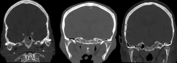
Figure 1. Coronal reconstruction of computed tomography demonstrating degenerative changes in the temporomandibular joint.
The sample was divided into three groups of seven participants each. Matching was pre‑specified in the study protocol and performed manually by an independent investigator who did not participate in the clinical evaluations. The matching variables included age (maximum difference of ±3 years), sex, baseline pain intensity assessed on a visual analog scale (VAS), and baseline mouth opening (maximum difference of ±3 mm).
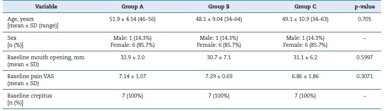
This procedure ensured clinical comparability among the groups prior to the interventions and reduced the risk of selection bias (Table 1).
Table 1. Baseline characteristics of patients by treatment group. Demographic and clinical variables (mean ± standard deviation, range, and frequency) for Groups A, B, and C at baseline, with corresponding p-values.
SD – standard deviation.
Each group received different therapeutic modalities: Group A received intra-articular infiltration of ozone-activated platelet-rich plasma (PRP-O3), Group B received a stabilization splint, and Group C received intra-articular infiltration of PRP-O3 combined with a stabilization splint. Intra-articular infiltration was performed in three sessions at 15-day intervals.
A wash‑out period of 7 days was established before the start of the intervention, during which the use of analgesics and anti‑inflammatory drugs was prohibited.
All clinical evaluations and interventions were performed by the same team of specialists following standardized protocols, thus minimizing variability and reducing the risk of bias related to clinician contact.
Peripheral venous blood (20 mL) was collected under minimal tourniquet application (<1 minute) into sterile heparinized tubes (30 IU/mL) to prevent coagulation and preserve platelet integrity. Samples were processed using an open technique under aseptic conditions. Double centrifugation was performed in a benchtop centrifuge (Eppendorf 5702R, rotor radius 15 cm). The first centrifugation was performed at 200 x g (1,130 rpm) for 10 minutes. The obtained plasma was carefully removed with a pipette, avoiding turbulence, and subjected to a second centrifugation at 100 x g (800 rpm) for 15 minutes.
PRP was obtained after discarding 0.5 mL of the upper fraction. Baseline platelet counts averaged 225,000 ± 40,000/μL, while PRP samples reached 880,000 ± 130,000/μL, representing na approximately 3.5‑fold increase compared to baseline. The PRP obtained was characterized as leukocyte-poor (<1,000/μL).
Ozone was generated using a medical-grade ozone generator (Ozonosan Photonic 101, Hansler, Germany), calibrated according to the manufacturer’s specifications. The concentration of 30 μg/mL was verified with the in‑line photometric system of the device, and manipulation time was strictly limited to less than 2 minutes to minimize ozone reactivity. For activation, a three‑way stopcock was used to connect one syringe containing PRP and another containing ozone. The syringes were alternately discharged into each other through the stopcock for one minute, ensuring homogeneous mixing at a 1:1 PRP–ozone ratio.
Patients were placed in a reclined position with maximum mouth opening. Skin antisepsis was ensured with 10% povidone‑iodine.
Anatomical references included the line between the tragus and the lateral canthus, with the insertion point located approximately 1 cm anterior to the tragus and 0.5 cm inferior in the vertical plane. All patients presented with bilateral osteoarthritis and received bilateral injections. The total amount injected per patient was 2 mL per session (1 mL per joint).
In Groups B and C, patients were instructed to use a rigid acrylic stabilization splint fabricated for the maxillary arch.
The appliance was designed with a flat occlusal scheme and canine guidance. It increased the vertical dimension by approximately 2 mm and was periodically adjusted to maintain uniform contacts in centric relation. Patients wore the splint exclusively at night for 12 consecutive weeks, with biweekly follow‑up visits scheduled to verify comfort, perform occlusal adjustments, and monitor adherence.
The variables studied were age, mouth opening (maximum painless incisal opening measured in mm with a calibrated vernier caliper), and pain (evaluated using the VAS).
Mouth opening and pain were assessed at baseline, 4 weeks, and 3 months. A trained and previously calibrated professional who was blinded to the patients’ group allocation measured these variables. Analyses were performed at the individual patient level.
All patients were systematically monitored for potential adverse events related to intra-articular infiltration and ozone manipulation. The following complications were specifically assessed: transient pain flare, swelling, hematoma, infection, transient facial nerve symptoms, vasovagal reactions, and need for rescue medication. Adverse events would be recorded at each follow-up visit, but none were observed during the study period.
Data processing was performed using descriptive statistics. Age was analyzed using one‑way analysis of variance (ANOVA), and when normality assumptions were not met, the non‑parametric Kruskal–Wallis test was applied. For continuous clinical variables (mouth opening and pain), a repeated‑measures ANOVA was used, with post hoc comparisons adjusted by Bonferroni correction. Assumptions of normality were checked using the Shapiro–Wilk test and graphical inspection of residuals, and sphericity was assessed with Mauchly’s test, acknowledging the limited power with n=7/ group. In addition to p‑values, effect sizes were reported as mean differences in change between groups with 95% confidence intervals, emphasizing magnitude and uncertainty rather than statistical significance alone. In case assumptions were not met, a more robust approach was considered, such as a linear mixed‑effects model with fixed effects for group and time and random intercepts for patients, or a non‑parametric strategy with transparent justification. A significance level of p<0.05 and a 95% confidence interval were applied in all analyses.
All patients signed an informed consent after being informed of the study characteristics and their right to withdraw at any time. Patient confidentiality and privacy were fully protected throughout the study. All clinical data were anonymized, and images were obtained with written informed consent. No personal identifiers or recognizable facial features were included in the manuscript or figures.
The study was reviewed and approved by the Ethics Committee of the Manuel Ascunce Domenech University Hospital and the Ethics Committee of the Faculty of Dentistry, University of Medical Sciences of Camagüey (Approval No. 12/2024).
All procedures involving human participants were conducted in accordance with the ethical standards of these institutional review boards and with the principles of the Declaration of Helsinki.17
Results
The groups had mean ages of 51.9, 48.1, and 49.1, respectively, indicating similar mean values, with no statistically significant differences (p=0.705) (Table 1). These findings confirm that the groups were comparable in terms of patient age, thereby excluding this variable as a confounding factor in subsequente results.
Baseline mouth opening values were similar among the groups (A: 32.9 mm; B: 30.7 mm; C: 31.1 mm), with a progressive increase over time. At three months, Group C achieved the highest mean (44.9 mm), surpassing Group A (38.6 mm) and Group B (33.7 mm) (Figure 2). Table 2 presents the descriptive statistics for each group and time point, including mean ± SD and change from baseline.
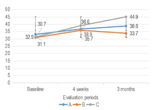
Figure 2. Analysis of mouth opening.
Table 2. Mouth opening in TMJ‑OA patients. Mean values (± standard deviation) for Groups A, B, and C at baseline, 4 weeks, and 3 months, with change from baseline.

SD – standard deviation.
Assumptions of normality were assessed using the Shapiro–Wilk test and graphical inspection of residuals, while sphericity was evaluated with Mauchly’s test, acknowledging the limited statistical power with n=7 per group. Bivariate analysis revealed significant differences between groups (p=0.013). Post hoc comparisons indicated that, at three months, Group C exhibited significantly greater mouth opening than Group B (p=0.0005), while its difference from Group A did not reach statistical significance (p=0.0716). In addition to p‑values, effect sizes were reported as mean differences in change between groups with 95% confidence intervals, highlighting both the magnitude and the uncertainty of the observed effects.
These findings demonstrate overall improvement over time, with a more pronounced effect in patients treated with the combination of PRP‑O3 and occlusal splint therapy.
Baseline pain values were high and comparable across groups (A: 7.14; B: 7.29; C: 6.86). At four weeks, Groups A and C showed greater reductions, while Group B remained higher. At three months, Group C achieved the lowest mean pain score (1.8), followed by Group A (2.0) and Group B (3.14) (Figure 3). Table 3 presents the descriptive statistics (mean ± SD and change from baseline).
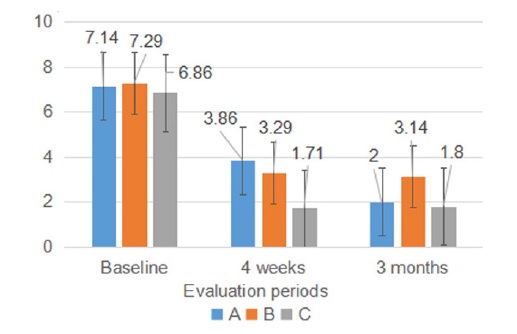
Figure 3. Analysis of pain
Table 3. Pain analysis in TMJ-OA patients. Mean values (± standard deviation) for Groups A, B, and C at baseline, 4 weeks, and 3 months, with change from baseline.

SD – standard deviation.
Normality was assessed with the Shapiro–Wilk test and residual plots, and sphericity with Mauchly’s test. Bivariate analysis confirmed significant differences between groups (p = 0.0009) and across time (p<0.001). Post hoc comparisons indicated that Group B had significantly higher pain than Group C at three months (p=0.0155), while other differences were not significant (p>0.05). Effect sizes with 95% confidence intervals highlighted the magnitude and uncertainty of these differences, indicating an overall reduction in pain, most pronounced in patients treated with PRP‑O3 associated with occlusal splint therapy.
Discussion
PRP has been consolidated as a multifactorial therapeutic strategy in tissue regeneration, acting not only through growth factors but also via adhesion molecules, immunomodulatory mediators, angiogenic factors, and pain modulators.
Its clinical application extends across various medical specialties, particularly dentistry, where it serves as a valuable tool for treating multiple lesions. Moreover, PRP-O3 has demonstrated high efficacy, especially when combined with conventional ozone therapy, thereby enhancing its therapeutic benefits.18
Studies have demonstrated that ozonation of heparinized plasma promotes platelet aggregation and, thus, enhances the release of growth factors, reinforcing the therapeutic efficacy of PRP in tissue regeneration.19, 20 Subsequently, this combination has been employed in different preparations for wound healing, cutaneous biostimulation, facial rejuvenation, and the treatment of osteoarthritis in various joints.11
The use of PRP-O3 in TMJ-OA has been reported in a study demonstrating its effectiveness and safety in 100 patients with this disease.16 The evaluation included pain, mouth opening, joint sounds, and the occurrence of adverse effects. Patients were divided into four groups based on the approach used: conservative treatment, PRP infiltration, ozone infiltration, and PRP-O3 infiltration. All groups received infiltrations every 15 days for three sessions, with 1 mL administered per infiltration.
Patients treated with PRP-O3 showed significantly better outcomes than the other groups. Several authors have reported satisfactory results with PRP in TMJ-OA.6, 7, 21 A study analyzing the combination of PRP injection with individualized comprehensive physiotherapy demonstrated clinical advantages compared to PRP alone in the treatment of temporomandibular joint osteoarthritis (TMJ-OA). In that study, although imaging findings were comparable between the two modalities and both groups exhibited progressive improvements in pain, mouth opening, and joint function, the combined approach resulted in a more pronounced reduction in pain and superior functional recovery.
These results suggest that integrating PRP with personalized physiotherapy enhances the therapeutic benefits in TMJ-OA.6
The efficacy of PRP infiltration has been evaluated against alternatives such as hyaluronic acid or saline solution. Most studies confirm that PRP provides sustained improvements in mandibular range of motion and pain reduction for up to one year after treatment. Nonetheless, comparable benefits have been reported across different therapeutic modalities.21
The flat stabilization device is the most widely used occlusal appliance in the management of temporomandibular disorders.
Typically fabricated for the upper arch, its primary objectives are to stabilize the joint, protect teeth, and reduce overload on the muscles and TMJ structures. When properly manufactured, it presents minimal adverse effects, which explains its broad clinical acceptance.22
A retrospective study analyzed 148 patients diagnosed with TMD to evaluate the efficacy of occlusal splint therapy on condylar position, joint morphology, and patient-reported outcomes. Results demonstrated significant improvement i condylar position, along with increased joint space and better disk positioning, while bone alterations were not clinically relevant.23
The efficacy of occlusal splints combined with PRP in patients with TMJ‑OA has been evaluated in China. After six months of treatment, significant reductions in pain and increases in maximum mouth opening were observed. These findings are consistent with the present study’s results and provide evidence that the combination of splint therapy and PRP can improve both pain symptoms and mandibular function in patients with TMJ‑OA.1 Moreover, when PRP is activated with ozone, the results suggest a more comprehensive and complementary therapeutic approach compared to splint therapy alone.
This study presents certain limitations that must be considered.
The small sample size and single-center design limit the generalizability of the findings, and the threemonth follow-up period prevents assessment of long-term efficacy. Moreover, the absence of blinding may introduce bias in pain perception and clinical evaluation. Nevertheless, patients were allocated based on age and clinical characteristics to ensure initial group similarity and reduce the risk of selection bias. Future studies should aim to expand the sample size and incorporate auxiliary diagnostic methods to evaluate articular cartilage regeneration, thereby providing more comprehensive evidence on the long-term outcomes of treatment.
Conclusions
The purpose of this study was to evaluate the efficacy of PRP-O3 combined with occlusal splint therapy in patients with TMJ-OA, and to present this combination as an innovative therapeutic modality, since no prior reports were found in the consulted literature.
Findings from previous investigations demonstrated that ozone acts as an excellent activator of PRP, with beneficial effects in degenerative joint disease. The results of the presente study confirm that intra-articular infiltration of PRP-O3 combined with occlusal splint therapy is associated with significantly superior outcomes in the recovery of mouth opening and pain relief compared with either PRP-O3 or occlusal splint therapy alone.
References
1. Wu CB, Sun NN, Zhang D, Wang Q, Zhou Q. Efficacy analysis of splint combined with platelet-rich plasma in the treatment of temporomandibular joint osteoarthritis. Front Pharmacol. 2022;13:996668.
2. Valesan LF, Da-Cas CD, Réus JC, Denardin ACS, Garanhani RR, Bonotto D, et al. Prevalence of temporomandibular joint disorders: a systematic review and meta-analysis. Clin Oral Investig. 2021;25(2):441-53.
3. Lee YH, Park HK, Auh QS, Nah H, Lee JS, Moon HJ, et al. Emerging potential of exosomes in regenerative medicine for temporomandibular joint osteoarthritis. Int J Mol Sci. 2020;21(4):1541.
4. Alqutaibi AY, Alhammadi MS, Hamadallah HH, Altarjami AA, Malosh OT, Aloufi AM, et al. Global prevalence of temporomandibular disorders: a systematic review and meta-analysis. J Oral Facial Pain Headache. 2025 Jun;39(2):48-65.
5. Al-Ani Z. Temporomandibular joint osteoarthrosis: a review of clinical aspects and management. Prim Dent J. 2021;10(1):132-40.
6. Liu SS, Xu LL, Fan S, Lu SJ, Jin L, Liu LK, et al. Effect of platelet-rich plasma injection combined with individualised comprehensive physical therapy on temporomandibular joint osteoarthritis: a prospective cohort study. J Oral Rehabil. 2022;49(2):150-9.
7. Derwich M, Mitus-Kenig M, Pawlowska E. Mechanisms of action and efficacy of hyaluronic acid, corticosteroids, and platelet-rich plasma in the treatment of temporomandibular joint osteoarthritis: a systematic review. Int J Mol Sci. 2021;22(14):7405.
8. Yuan W, Wu Y, Huang M, Zhou X, Liu J, Yi Y, et al. A new frontier in the treatment of temporomandibular joint osteoarthritis: exosome-based therapeutic strategy. Front Bioeng Biotechnol. 2022;10:1074536.
9. Ramírez Suarez R, Quesada Leyva L, Nicolau Pestana E, Mosquera Betancourt G. Estabilidad farmacológica del plasma rico en plaquetas activado con ozono. Rev Cubana Hematol Inmunol Hemoter [Internet]. 2025 [cited 2026 Jun 4];41. Available from: https://revhematologia.sld.cu/index.php/hih/article/view/2162
10. Pretorius J, Habash M, Ghobrial B, Alnajjar R, Ellanti P. Current status and advancements in platelet-rich plasma therapy. Cureus. 2023;15(10):e47176.
11. Schwartz Tapia A, Martínez Sánchez G, Re L. Factores de crecimiento derivados de plaquetas y sus aplicaciones en medicina regenerativa. Potencialidades del uso del ozono como activador. Revista Española de Ozonoterapia [Internet]. 2011 [cited 2026 Jun 4];1(1):54-73. Available from: https://dialnet.unirioja.es/descarga/articulo/3658622.pdf
12. Ramírez-Suarez R, Mosquera-Betancourt G, Tan-Suarez N, Morales-Paz YR, Morales-Basulto R. Validación de la infiltración intraarticular de plasma rico en plaquetas ozonizado en osteoartritis temporomandibular. Arch Méd Camagüey [Internet]. 2025 [cited 2026 Jun 4];29:e10966. Available from: https://revistaamc.sld.cu/index.php/amc/article/view/10966
13. Bhattacharjee B, Bera RN, Verma A, Soni R, Bhatnagar A.Efficacy of arthrocentesis and stabilization splints in the management of temporomandibular joint disc displacement disorder without reduction: a systematic review and meta-analysis. J Maxillofac Oral Surg. 2023;22(1):83-93.
14. Li DTS, Luo LY, Li KY, Su YX, Durham J, Leung YY. Early arthrocentesis for temporomandibular joint arthralgia: a superiority trial. Int Dent J. 2024;74(6):1362-70.
15. Wang S, Wang Z, Zhou G. Efficacy of added splint therapy after arthrocentesis vs arthrocentesis alone in the management of temporomandibular joint disorders: a systematic review and meta-analysis. Quintessence Int. 2024;55(10):824-33.
16. Ramírez Suarez R, Mosquera Betancourt G, Tan Suárez N, Pagés Morales N. Efecto y seguridad del plasma rico en plaquetas ozonizado en pacientes con osteoartritis temporomandibular. Rev Asoc Odontol Argent. 2025;113(2):e1130813.
17. World Medical Association. WMA Declaration of Helsinki – Ethical Principles for Medical Research Involving Human Participants [Internet]. 2024 Oct 19 [cited 2026 Jun 4]. Available from: https://www.wma.net/policies-post/wma-declaration-of-helsinki/
18. Wu WS, Chen LR, Chen KH. Platelet-rich plasma (PRP):molecular mechanisms, actions and clinical applications in human body. Int J Mol Sci. 2025;26(21):10804.
19. Valacchi G, Bocci V. Studies on the biological effects of ozone:10. Release of factors from ozonated human platelets. Mediators Inflamm. 1999;8(4-5):205-9.
20. Re L, Martínez-Sánchez G, Perez-Davison G, Sirito M. Role of ozone/oxygen in fibroblast growth factor activation. Discovering the facts. Int J Ozone Ther. 2010;9(2):55-8.
21. Haddad C, Zoghbi A, El Skaff E, Touma J. Platelet-rich plasma injections for the treatment of temporomandibular joint disorders: a systematic review. J Oral Rehabil. 2023;50(11):1330-9.
22. Albagieh H, AlWazzan AK, Alhelal FA, Alem MF, Albaiz AM, Aloraini TK, et al. Effectiveness of occlusal splints in the management of temporomandibular disorders: comparisons of treatment approaches and digital versus conventional fabrication techniques. Cureus. 2025;17(1):e77451.
23. Mathew A, Cr V, Ka A, Goswami D, Antony T, Bharat R. Effectiveness of occlusal splint therapy in moderating temporomandibular joint disorders with joint displacement: a retrospective analysis using cone beam computed tomography. Cureus. 2024;16(3):e57300.
Reynier Ramírez
E-mail address: reynierramirez93@gmail.com
CRediT authorship contribution statement
Reynier Ramírez: Conceptualization, Data curation, Formal analysis, Investigation, Methodology, Project administration, Supervision, Validation, Visualization, Writing – original draft, Writing - review & editing. Yoan R. Arango: Data curation, Formal analysis, Validation, Visualization, Writing – original draft.
Ethical disclosures
Protection of human and animal subjects. The authors declare that the procedures followed were in accordance with the regulations of the relevant clinical research ethics committee and with those of the Code of Ethics of the World Medical Association (Declaration of Helsinki).
Confidentiality of data. TThe authors declare that they have followed their work center protocols on access to patient data and for its publication.
Right to privacy and informed consent. The authors have obtained the written informed consent of the patients or subjects mentioned in the article. The corresponding author is in possession of this document.
Conflict of interest
The authors have no conflicts of interest to declare.
Acknowledgments
This study received no external funding. An AI language tool was used to assist with the Portuguese translation of the abstract. The translated text was reviewed and verified for accuracy by a bilingual author. The authors accept full responsibility for the accuracy and appropriateness of the translated content.
1646-2890/© 2026 Sociedade Portuguesa de Estomatologia e Medicina Dentária. Published by SPEMD.
This is an open access article under the CC BY-NC-ND license (http://creativecommons.org/licenses/by-nc-nd/4.0/).