

Revista Portuguesa de Estomatologia, Medicina Dentária e Cirurgia Maxilofacial
SPEMD - SPEMD - Revista Portuguesa de Estomatologia Medicina Dentária e Cirurgia Maxilofacial | 2026 | 67 (2) | 48-55
Original research
Gap arthroplasty outcomes in temporomandibular joint ankylosis: a retrospective study
Desfechos da artroplastia de gap na anquilose pediátrica da articulação temporomandibular: um estudo retrospetivo
a Department of Oral and Maxillofacial Surgery, Khyber College of Dentistry, Peshawar, Pakistan
b Naseer Teaching Hospital, Peshawar, Pakistan
c Kabir Medical College, Peshawar, Pakistan
d Khyber College of Dentistry, Peshawar, Pakista
Qazi Jawad Hayat - Qazijawadhayat@gmail.com
Article Info
Rev Port Estomatol Med Dent Cir Maxilofac
Volume - 67
Issue - 2
Original research
Pages - 48-55
Go to Volume
Article History
Received on 02/03/2026
Accepted on 23/05/2026
Available Online on 30/06/2026
Keywords
Original Research
Gap arthroplasty outcomes in temporomandibular joint ankylosis: a retrospective study
Desfechos da artroplastia de gap na anquilose pediátrica da articulação temporomandibular: um estudo retrospetivo
Muhammad Owais Khan1 0009-0001-4985-9735
Hadia Shehzad2 0009-0006-7255-1786
Asra Hayat3 0009-0000-3761-1604
Qazi Jawad Hayat4,* 0009-0009-6879-8884
1 Department of Oral and Maxillofacial Surgery, Khyber College of Dentistry, Peshawar, Pakistan
2 Naseer Teaching Hospital, Peshawar, Pakistan
3 Kabir Medical College, Peshawar, Pakistan
4 Khyber College of Dentistry, Peshawar, Pakistan
Article history:
Received 2 March 2026
Accepted 23 May 2026
Available online 24 June 2026
Abstract
Objectives: To evaluate postoperative maximal inter-incisal opening and facial nerve injury following gap arthroplasty, and to assess the influence of Sawhney classification on these outcomes.
Methods: Data from 103 patients (Sawhney Class II, n=28; Class III, n=75) undergoing gap arthroplasty were reviewed. Postoperative maximal inter-incisal opening and facial nerve weakness were recorded at outpatient follow-up (mean 2.26 months, SD 1.42; range 1–8 months). All continuous variables showed non-normal distribution (Shapiro-Wilk p<0.05); the Mann–Whitney U test and Fisher’s exact test were applied. A Kruskal-Wallis test was used for age-stratified mouth opening analysis. Preoperative maximal inter-incisal opening was not available, precluding pre-post comparison.
Results: Mean age was 12.26 ± 3.35 years. Falls were the leading cause (80.6%). Mean postoperative maximal inter-incisal opening was 28.42 ± 7.09 mm (range 5–42 mm); 71.8% of patients achieved ≥25 mm. No significant difference in mouth opening was observed between Class II (28.43 ± 5.17 mm) and Class III (28.45 ± 7.71 mm; p=0.991). Age-stratified analysis showed progressively higher postoperative mouth opening in older patients (5–9 years: 24.8 mm; 10–13 years: 28.9 mm; 14–17 years: 30.4 mm; Kruskal-Wallis p=0.038). Facial nerve weakness occurred in 21.4% of patients overall (Class II: 21.4%; Class III: 21.3%), with no association with Sawhney classification (p=1.000).
Conclusion: Gap arthroplasty resulted in satisfactory short-term mouth opening. Sawhney classification did not influence maximal inter-incisal opening or facial nerve injury risk. Postoperative mouth opening was positively associated with patient age, consistent with expected developmental growth patterns. The absence of preoperative data and short follow- up are primary limitations. Prospective studies with long-term follow-up are recommended.
Keywords: Ankylosis, Arthroplasty, Facial nerve injuries,Mouth opening,Retrospective studies,Temporomandibular joint disorders
Resumo
Objetivos: Avaliar a abertura interincisal máxima pós-operatória e a ocorrência de lesão do nervo facial após artroplastia de gap e analisar a influência da classificação de Sawhney nesses desfechos.
Métodos: Foram analisados dados de 103 pacientes (Classe II de Sawhney, n=28; Classe III, n=75) submetidos a artroplastia de gap. A abertura interincisal máxima pós-operatória e a fraqueza do nervo facial foram avaliadas no seguimento em ambulatório (média 2,26 meses, DP 1,42; intervalo 1–8 meses). Todas as variáveis contínuas apresentaram distribuição não normal (Shapiro-Wilk p<0,05); foram utilizados o teste de Mann–Whitney U e o teste exato de Fisher. O teste de Kruskal-Wallis foi utilizado para análise estratificada por faixa etária. A abertura interincisal máxima pré-operatória não estava disponível.
Resultados: A idade média foi de 12,26 ± 3,35 anos. As quedas foram a principal causa (80,60%). A abertura interincisal máxima média pós-operatória foi de 28,42 ± 7,09 mm; 71,80% dos pacientes atingiram ≥25,00 mm. Não houve diferença significativa entre as Classes II (28,43 ± 5,17 mm) e III (28,45 ± 7,71 mm; p=0,99). A análise estratificada por faixa etária demonstrou abertura interincisal máxima progressivamente maior nos pacientes mais velhos (5–9 anos: 24,80 mm; 10–13 anos: 28,90 mm; 14–17 anos: 30,40 mm; Kruskal-Wallis p=0,04). A fraqueza do nervo facial ocorreu em 21,36% dos casos, sem associação à classificação de Sawhney (p=1,00).
Conclusões: A artroplastia de gap apresentou resultados satisfatórios a curto prazo. A classificação de Sawhney não influenciou os desfechos avaliados. A abertura interincisal máxima pós-operatória apresentou associação positiva com a idade, consistente com os padrões esperados de crescimento e desenvolvimento. São necessários estudos prospetivos com seguimento prolongado.
Palavras-chave: Anquilose,Artroplastia,Traumatismos do nervo facial,Abertura bucal, Estudos retrospetivos,Transtornos da articulação temporomandibular
Introduction
Temporomandibular joint (TMJ) ankylosis is a debilitating condition characterized by fusion of the mandibular condyle to the glenoid fossa, resulting in severe restriction of mandibular movements. This condition compromises mastication, speech, oral hygiene, facial aesthetics, and psycho-social well-being. When ankylosis develops during the growth period, the consequences are more profound, often leading to mandibular hypoplasia, retrognathism, facial asymmetry, malocclusion, and, in severe cases, obstructive sleep apnea.1, 2
Trauma remains the most common cause of TMJ ankylosis, particularly in children, where untreated condylar fractures frequently progress to fibrous or bony fusion.3 The young population is especially vulnerable due to the high osteogenic potential of the condyle and the delicate anatomical structure of the joint.4 Ankylosis may be fibrous, bony, or mixed; complete bony ankylosis represents the most severe form and typically requires surgical intervention.5 - 7
The most important clinical manifestation of TMJ ankylosis is limitation of mouth opening, measured as maximal inter- incisal opening (MIO). Surgical management aims to restore mandibular mobility, prevent recurrence, and preserve surrounding structures, particularly the facial nerve (CN VII).
Facial nerve dysfunction is one of the most concerning complications of TMJ surgery. Neuropraxia resulting from stretching and retraction of the frontal and zygomatic branches during the preauricular approach is the most common mechanism of injury.8
Gap arthroplasty remains a widely accepted surgical technique owing to its relative simplicity and reliable short-term functional outcomes when combined with aggressive postoperative physiotherapy.9, 10 However, there is a lack of studies, particularly in South Asian pediatric populations, on whether the severity of ankylosis, as defined by the Sawhney classification, independently predicts postoperative MIO or facial nerve morbidity following gap arthroplasty11 - 14
The present study aimed to evaluate postoperative MIO and the frequency of facial nerve injury following gap arthroplasty, and to determine whether Sawhney classification influences these short-term outcomes.
Material and Methods
This retrospective observational study was conducted at the Department of Oral and Maxillofacial Surgery, Khyber College of Dentistry, Peshawar, Pakistan, from April 2022 to June 2024 (27 months). Data were collected consecutively from patients who underwent gap arthroplasty and presented to the outpatient department (OPD) for postoperative follow-up during the study period. There were no fixed pre-scheduled follow-up intervals; patients were assessed at whichever OPD visit occurred within the study period.
This study was approved by the Institutional Ethics Committee of Khyber College of Dentistry, Peshawar (Reference No.: 3010/RRB/KCD, dated 09/09/2021), in accordance with the Declaration of Helsinki. Written and verbal informed consent were obtained from all patients or their legal guardians (for patients under 18 years of age) prior to inclusion, including consent for use of anonymized clinical data in research. All patient data were anonymized prior to analysis, and no identifying information is included in this report. Confidentiality was maintained throughout.
Using the WHO sample size calculator, assuming a frequency of facial nerve damage of 13.02%,1 with an absolute precision of 6.5% at a 95% confidence interval, the calculated sample size was 103 patients. Sampling was non-probability consecutive.
Patients with radiologically confirmed TMJ ankylosis (Sawhney Class II or III on CT scan or CBCT) who underwent gap arthroplasty at the study center during the study period were included. The original eligibility criterion was age 2–30 years, set to capture the range that most commonly presents bony ankylosis following condylar injury: the minimum age of 2 years reflects the earliest at which bony ankylosis can be radiologically confirmed and surgically managed; the maximum age of 30 years was set to capture young adults in whom untreated childhood ankylosis may first present to tertiary care, consistent with the inclusion ranges of comparable regional studies.11,12 The actual enrolled sample comprised patients aged 5–17 years, reflecting the pediatric-predominant patient population at this center. Sawhney Class I (fibrous, typically non-surgical) and Class IV (not encountered during the study period) were excluded. Patients with systemic joint or bone disorders, recurrent re-ankylosis, neuromuscular disorders affecting facial movement, or incomplete clinical records were also excluded. Radiological classification was established using CT scan or CBCT: Class II denotes lateral bony ankylosis with partial fusion; Class III denotes complete bony ankylosis with no recognizable condyle.
All gap arthroplasty procedures were performed by the same surgical team under the direct supervision of the consultant surgeon. Under general anesthesia, the TMJ was exposed via a standard preauricular approach. Two horizontal osteotomy cuts were made, a bone wedge was removed, and a minimum gap of 1 cm was created between the glenoid fossa and the ramus. No interposition material was used. Ipsilateral coronoidectomy was performed where clinically indicated (in cases of clinically assessed coronoid hypertrophy contributing to restricted mouth opening). The number of patients who underwent concomitant coronoidectomy was inconsistently documented in the retrospective dataset and could not be reliably extracted; this is acknowledged as a limitation affecting the interpretation of the MIO analysis, as coronoidectomy may represent an uncontrolled covariate. Wound closure was achieved in layers.
Early active jaw-opening exercises were advised for all patients and reinforced at OPD visits. A standardized protocol specifying start day, frequency, duration, exercise devices, and adherence monitoring was inconsistently documented in this retrospective cohort; this is acknowledged as a limitation affecting reproducibility.
The primary outcome measures were: (1) postoperative MIO measured as the inter-incisal distance between upper and lower central incisors using a calibrated ruler in millimeters, recorded by the postgraduate resident at the follow-up visit; and (2) facial nerve weakness assessed clinically by observing loss or weakness of facial expression in the fronto-zygomatic distribution on the operated side, recorded as a binary outcome (present/absent). The House-Brackmann grading scale was not applied; this is acknowledged as a limitation. Preoperative MIO was not recorded in this retrospective dataset, precluding pre-post comparison; this is stated as a primary limitation.
Data were analyzed using SPSS version 27. Normality was assessed using Kolmogorov-Smirnov and Shapiro-Wilk tests for all continuous variables: age, K-S p=0.000, Shapiro-Wilk p=0.000; duration after surgery, K-S p=0.000, Shapiro-Wilk p=0.001; postoperative MIO, K-S p=0.000, Shapiro-Wilk p=0.001.
All three variables showed non-normal distribution (all p<0.05), justifying non-parametric tests. The Mann-Whitney U test was used to compare postoperative MIO between Sawhney classes and between age groups (Kruskal-Wallis for three age groups).
Fisher’s exact test was used for categorical associations. A p-value of ≤0.05 was considered statistically significant.
Results
A total of 103 patients were included (Table 1). The enrolled sample comprised patients aged 5–17 years, and more males (n=71) than females (n=32). The most common cause of TMJ ankylosis was falls (80.6%), followed by road traffic accidents (14.6%). Ankylosis was unilateral in 73.8% of cases and bilateral in 26.2%. Sawhney Class III was the predominant classification (72.8%), and Class II accounted for 27.2% of patients. The mean follow-up duration was 2.26 months (SD 1.42).
Table 1. Descriptive statistics and demographic characteristics (n=103)
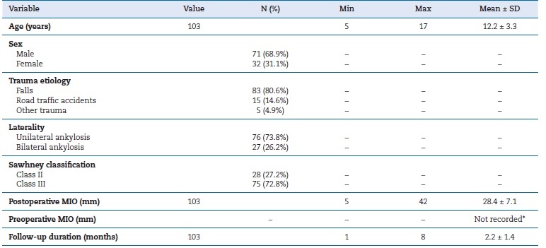
MIO – Maximal inter-incisal opening. * Preoperative MIO was not recorded in this retrospective dataset.
The overall mean postoperative MIO was 28.42 ± 7.09 mm. The lowest recorded value was 5 mm, representing severe functional limitation well below the 25 mm functional threshold, noted in a single patient (Sawhney Class III) assessed at one month postoperatively. This early assessment may not reflect the final functional outcome, and the value likely represents incomplete recovery at the time of the single recorded follow-up visit. Twenty-nine patients (28.2%) did not achieve the 25 mm functional threshold at their recorded follow-up visit. Class-wise mean postoperative MIO was 28.43 ± 5.17 mm for Class II and 28.45 ± 7.71 mm for Class III (Table 2). The Mann-Whitney U test revealed no statistically significant difference between groups (p=0.991; Figure 1). Seventy-four patients (71.8%) achieved a postoperative MIO ≥25 mm, a threshold associated with functional adequacy for mastication and oral hygiene. Since preoperative MIO data were unavailable in this retrospective dataset, no pre-post comparison was made.
Table 2. Postoperative MIO by Sawhney classification

MIO – Maximal inter-incisal opening. * Mann-Whitney U test
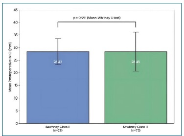
Figure 1. Bar graph showing the mean postoperative maximal inter-incisal opening (MIO) in millimeters by Sawhney classification. Error bars represent ± 1 standard deviation (Class II: 28.43 ± 5.17 mm; Class III: 28.45 ± 7.71 mm). No statistically significant difference was observed (Mann-Whitney U, p=0.991).
Table 3 presents age-stratified postoperative MIO. A Kruskal-Wallis test indicated a statistically significant difference across age groups (p=0.038; Figure 3), with older patients achieving higher postoperative MIO. This pattern is consistente with developmental growth and expected normative increases in MIO with age, and does not represent a difference in surgical efficacy per se.
Table 3. Postoperative MIO stratified by age group

MIO – Maximal inter-incisal opening. * Kruskal-Wallis test across three age groups. Age groups defined by developmental stage (primary/early
mixed dentition; late mixed/early permanent dentition; adolescent).
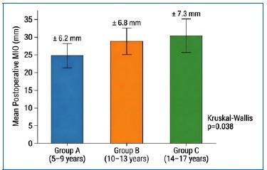
Figure 3. Bar graph showing mean postoperative MIO stratified by age group. Group A (5–9 years): 24.8 ± 6.2 mm; Group B (10–13 years): 28.9 ± 6.8 mm; Group C (14–17 years): 30.4 ± 7.3 mm. Error bars represent ± 1 SD. Kruskal-Wallis p=0.038.
Table 4 presents the covariate comparison between Sawhney Class II and Class III patients. No statistically significant differences were identified in mean age (p=0.41), sex distribution (p=0.32), unilateral/bilateral status (p=0.61), or etiology (p=1.000) between the two classes, indicating that the groups were broadly comparable with respect to available covariates. Facial nerve weakness in the fronto-zygomatic distribution occurred in 21.4% of patients (Table 5; Figure 2). Class-specific rates were nearly identical, at 21.4% for Class II and 21.3% for Class III. Fisher’s exact test showed no statistically significant association between Sawhney classification and facial nerve weakness (p=1.000). The weakness represented paresis consistente with neuropraxia from preauricular retraction; House-Brackmann grading was not performed, and the recovery timeline was not recorded.
Table 4. Covariate comparison between Sawhney Class II and Class III

† Mann-Whitney U test. ‡ Fisher’s exact test. No statistically significant covariate differences were identified between the two classes.
Table 5. Facial nerve weakness by Sawhney classification

* Fisher’s exact test (2-sided). Weakness assessed as a binary outcome (present/absent) at a single postoperative time point.
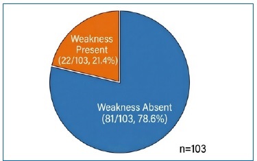
Figure 2. Overall distribution of postoperative facial nerve weakness following gap arthroplasty. Pie chart showing the proportion of patients with weakness present (22/103, 21.4%) versus absent (81/103, 78.6%) across the entire cohort (n=103). Class-specific rates were nearly identical: Class II 6/28 (21.4%); Class III 16/75 (21.3%). No statistically significant association with Sawhney classification was observed (Fisher’s exact test p=1.000).
Discussion
This retrospective observational study of 103 pediatric and adolescent patients (aged 5–17 years) treated with gap arthroplasty for TMJ ankylosis at a single tertiary center provides short-term postoperative outcome data stratified by Sawhney classification. The principal findings are: (1) the mean postoperative MIO was 28.42 mm at a mean follow-up of 2.26 months; (2) Sawhney classification did not significantly influence postoperative MIO (p=0.991); (3) facial nerve weakness occurred in 21.4% of patients and was not associated with Sawhney classification (p=1.000); and (4) older patients (14–17 years) achieved significantly higher postoperative MIO than younger patients (5–9 years) across all Sawhney classes.
The mean postoperative MIO of 28.42 mm at a mean follow- up of 2.26 months is consistent with, although lower than, outcomes from comparable longer-term series. Bhatt et al. reported a mean mouth opening of 33.2 mm in the gap arthroplasty group at a mean follow-up of 27.6 months in a mixed adult-pediatric cohort; our shorter mean follow-up of 2.26 months and exclusively pediatric sample likely account for the lower value.15 - 17 Park et al. reported a mean MIO exceeding 35 mm at a follow-up of more than six years, further illustrating the trajectory of ongoing MIO improvement with time and physiotherapy.9 Roychoudhury et al. reported satisfactory functional restoration following gap arthroplasty with coronoidectomy and active jaw exercises, irrespective of patient age.2 The lower MIO observed in the present cohort is therefore attributable to the considerably shorter follow-up period rather than to an inferior surgical technique. Despite this, 71.8% of patients achieved a postoperative MIO ≥25 mm, a threshold widely regarded as compatible with functional mastication and oral hygiene.
The absence of a statistically significant difference in postoperative MIO between Sawhney Class II (28.43 mm) and Class III (28.45 mm) patients (p=0.991) is clinically noteworthy.
Although Class III ankylosis represents more extensive bony fusion requiring more aggressive resection, this did not translate into inferior short-term functional recovery. This is consistent with Al-Moraissi et al.’s systematic review and meta-analysis, which reported that ankylosis severity did not significantly affect surgical outcomes following gap arthroplasty.18, 19 The covariate comparison (Table 4) demonstrates no significant differences in age, sex, laterality, or etiology between Class II and Class III patients in this cohort, strengthening the validity of this comparison. These findings suggest that adequate gap creation and structured postoperative physiotherapy may be more important determinants of short-term MIO than the radiological extent of the ankylotic mass. However, given the absence of preoperative MIO data and the short follow-up, this inference must be interpreted cautiously.
The age-stratified analysis (Table 3) revealed a statistically significant progressive increase in postoperative MIO from Group A (5–9 years: 24.8 mm) to Group C (14–17 years: 30.4 mm; Kruskal-Wallis p=0.038). This gradient is consistent with developmental maturation: MIO normative values increase with age as the mandible grows and musculature develops. Future studies should report age-stratified MIO to permit meaningful comparisons with age-matched normative data and with other published cohorts.
The 21.4% incidence of facial nerve weakness (fronto-zygomatic distribution) falls within the published range of 4–40% for the preauricular approach.20, 21 Bhatt et al. reported facial nerve dysfunction in 13.6% of patients undergoing gap arthroplasty via the preauricular approach at a mean follow-up of 27.6 months. Liu et al. reported a 12.5–32% incidence using the endaural approach. Weinberg and Kryshtalskyj reported comparable rates with the preauricular approach. The near-identical rates between Sawhney Class II (21.4%, 6/28) and Class III (21.3%, 16/75) strongly suggest that facial nerve injury in this series is primarily related to surgical manipulation and retraction rather than the size of the ankylotic mass.
Understanding the mechanism of facial nerve injury is importante for surgical planning and patient counseling. The frontal and zygomatic branches of CN VII run superficially within the temporo-parietal fascia in the preauricular region. During the preauricular approach, these branches are susceptible to neuropraxia, which is a reversible conduction block, from retraction, compression, or inadvertent stretching. Risk factors include: the extent of dissection required for access, the duration of retraction, inadvertent hemostatic suturing, and the surgeon’s experience. The absence of a mass-size effect in our data is consistent with retraction-mediated injury rather than injury from proximity to or resection of the ankylotic mass. Surgical strategies that have been demonstrated to reduce facial nerve morbidity include: meticulous identification of the frontal branch prior to retraction using anatomical landmarks (inter-tragal notch to lateral eyebrow), use of intraoperative facial nerve monitoring where available, limiting retraction duration and force, maintaining the plane of dissection deep to the temporoparietal fascia, and careful layer-by-layer wound closure.13 Formal prospective documentation of retraction duration and intraoperative nerve monitoring status would enable future studies to quantify the contribution of these factors.
The study population was predominantly male (68.9%), consistent with the higher incidence of trauma-related TMJ ankylosis in males in this demographic, as reported by Aleem and Nagi.22, 23 The predominance of falls as the etiological factor (80.6%) is representative of the local pediatric population in Khyber Pakhtunkhwa, Pakistan, where unprotected falls from heights remain the leading cause of facial trauma in children.
The following limitations must be acknowledged. First, the sample size was calculated based on the expected frequency of facial nerve damage rather than on the expected difference in MIO between Sawhney classes; accordingly, the study may have been underpowered to detect a clinically meaningful difference in MIO between groups, and the null finding (p=0.991) should be interpreted with this caveat in mind. Second, the absence of preoperative MIO data is the most critical limitation; without baseline measurements, the magnitude of functional improvement cannot be quantified. Third, the mean follow-up of 2.26 months is insufficient to assess long-term outcomes or recurrence, which is the defining outcome criterion in TMJ ankylosis surgery. Fourth, facial nerve weakness was recorded as a binary outcome at a single postoperative time point without House-Brackmann grading, preventing characterization of severity or time to resolution. Fifth, a standardized physiotherapy protocol was not documented, limiting reproducibility. Sixth, individual physiotherapy adherence by Sawhney class was not recorded. Seventh, the number of patients who underwent concomitant coronoidectomy was not reliably documented in this retrospective dataset, precluding its inclusion as a covariate in the MIO analysis; this may represent an uncontrolled confound. Eighth, median and interquartile range values for MIO were not available from the data source. Future prospective studies with preoperative baseline recording, House-Brackmann grading, structured follow-up at 3, 6, 12, and 24 months, and formal physiotherapy documentation are strongly recommended.
Conclusions
In this retrospective observational cohort of pediatric and adolescente patients, gap arthroplasty achieved a mean postoperative MIO of 28.42 mm at a mean short-term follow-up of 2.26 months, with 71.8% of patients achieving a clinically functional mouth opening ≥25 mm. Sawhney classification did not significantly influence postoperative MIO or the risk of facial nerve injury. Age-stratified analysis demonstrated a significant increase in postoperative MIO with advancing age group, consistent with developmental growth patterns. Facial nerve weakness occurred in 21.4% of patients, consistent with published rates for the preauricular approach, and appeared related to surgical retraction rather than ankylosis severity.
These findings suggest that standardized surgical technique and early postoperative physiotherapy are important determinants of short-term functional recovery regardless of Sawhney class. The absence of preoperative baseline data, short follow-up, and lack of recurrence assessment are significant limitations, and hence, prospective studies addressing these gaps are necessary before stronger conclusions can bedrawn.24
References
1. Bhatt K, Roychoudhury A, Bhutia O, Pandey RM. Functional outcomes of gap and interposition arthroplasty in the treatment of temporomandibular joint ankylosis. J Oral Maxillofac Surg. 2014;72(12):2434-9.
2. Roychoudhury A, Parkash H, Trikha A. Functional restoration by gap arthroplasty in temporomandibular joint ankylosis: a report of 50 cases. Oral Surg Oral Med Oral Pathol Oral Radiol Endod. 1999;87(2):166-9.
3. Erol B, Tanrikulu R, Görgün B. A clinical study on maxillofacial fractures in children. J Craniomaxillofac Surg. 2004;32(3):163-7.
4. Vasconcelos BC, Bessa-Nogueira RV, Cypriano RV. Temporomandibular joint ankylosis. Braz J Otorhinolaryngol. 2008;74(1):34-8.
5. Al-Kayat A, Bramley P. A modified pre-auricular approach to the temporomandibular joint and malar arch. Br J Oral Surg. 1979;17(2):91-103.
6. Al-Moraissi EA, Wolford LM, Ellis E, Neff A. Does temporomandibular joint ankylosis severity affect surgical outcome? A systematic review and meta-analysis. J Craniomaxillofac Surg. 2020;48(5):453-61.
7. Zhang Y, He DM, Zhu M. Radiologic classification and surgical implications in temporomandibular joint ankylosis. J Oral Rehabil. 2020;47(6):709-16.
8. Weinberg S, Kryshtalskyj B. Facial nerve function following temporomandibular joint surgery using the preauricular approach. J Oral Maxillofac Surg. 1992;50(10):1048-51.
9. Park MW, Eo MY, Seo BY, Nguyen TTH, Kim SM. Gap arthroplasty with active mouth opening exercises using an interocclusal splint in temporomandibular joint ankylosis patients. Maxillofac Plast Reconstr Surg. 2019;41(1):28.
10. Liu F, Giannakopoulos H, Quinn PD, Granquist EJ. Retrospective study of facial nerve function following temporomandibular joint arthroplasty using the endaural approach. Craniomaxillofac Trauma Reconstr. 2015;8(2):88-93.
11. Sharma P, Saxena V, Bharadwaj M. Etiology and management trends in temporomandibular joint ankylosis. J Maxillofac Oral Surg. 2021;20(2):230-7.
12. Elgazzar RF, Abdelhady AI, Saad KA, Elshaal MA. Gap versus interpositional arthroplasty in treatment of temporomandibular joint ankylosis. J Oral Maxillofac Surg. 2020;78(5):811-18.
13. Singh V, Bhagol A, Dhingra R, Kumar P. Facial nerve injury following temporomandibular joint surgery: incidence and recovery. J Craniofac Surg. 2019;30(7):2140-4.
14. Sawhney CP. Bony ankylosis of the temporomandibular joint: follow-up of 70 patients treated with arthroplasty and acrylic spacer interposition. Plast Reconstr Surg. 1986;77(1):29-38.
15. Bhatt K, Roychoudhury A, Bhutia O, Pandey RM. Functional outcomes of gap and interposition arthroplasty in the treatment of temporomandibular joint ankylosis: a comparative study. Int J Oral Maxillofac Surg. 2014;43(11):1313-9.
16. Roychoudhury A, Srivastava A, Parkash H. Functional outcomes of gap arthroplasty in temporomandibular joint ankylosis: long-term evaluation. Int J Oral Maxillofac Surg. 2022;51(9):1178-84.
17. Khan M, Ullah H, Rahman AU. Long-term functional outcomes following surgical management of temporomandibular joint ankylosis. J Oral Maxillofac Surg. 2023;81(2):265-72.
18. Khatri A, Kalra P, Singh M. Influence of ankylosis type on postoperative mouth opening following gap arthroplasty. J Craniofac Surg. 2024;35(1):92-6.
19. Ahmed N, Ali K, Rehman B. Predictors of successful surgical outcome in temporomandibular joint ankylosis. Int J Oral Maxillofac Surg. 2024;53(1):45-52.
20. Wang H, Zhang J, Li Z. Complications associated with temporomandibular joint ankylosis surgery: a clinical analysis. Oral Surg Oral Med Oral Pathol Oral Radiol. 2019;128(4):347-52.
21. Patel A, Shah R, Desai R. Recovery pattern of facial nerve weakness following preauricular approach in TMJ surgery. J Craniofac Surg. 2021;32(6):2156-60.
22. Aleem B, Nagi A. Age and gender distribution in patients with temporomandibular joint ankylosis attending teaching hospitals of Lahore. Pak J Med Health Sci. 2023;17(10):859.
23. Gupta VK, Mehrotra D, Malhotra S, Kumar S, Agarwal GG, Pal US. An epidemiological study of temporomandibular joint ankylosis. Natl J Maxillofac Surg. 2012;3(1):25-30.
24. Gupta S, Gupta H, Jain S. Role of aggressive physiotherapy in preventing recurrence of temporomandibular joint ankylosis. J Maxillofac Oral Surg. 2022;21(4):1120-6.
Qazi Jawad Hayat
E-mail address: Qazijawadhayat@gmail.com
CRediT authorship contribution statement
Muhammad Owais Khan: Conceptualization, Data curation, Formal analysis, Investigation, Methodology, Writing – original draft, Writing – review & editing. Hadia Shehzad: Data curation, Investigation, Writing – review & editing. Asra Hayat: Data curation, Resources, Writing – review & editing. Qazi Jawad Hayat: Conceptualization, Methodology, Project administration, Supervision, Validation, Writing – original draft, Writing – review & editing.
Conflict of interest
The authors have no conflicts of interest to declare.
Ethical disclosures
Protection of human and animal subjects. The authors declare that the procedures followed were in accordance with the regulations of the relevant clinical research ethics committee and with those of the Code of Ethics of the World Medical Association (Declaration of Helsinki).
Confidentiality of data. The authors declare that no patient data appear in this article.
Right to privacy and informed consent. The authors have obtained the written informed consent of the patients or subjects mentioned in the article. The corresponding author is in possession of this document.
© 2026 Sociedade Portuguesa de Estomatologia e Medicina Dentária. Published by SPEMD.
This is an open access article under the CC BY-NC-ND license (http://creativecommons.org/licenses/by-nc-nd/4.0/).