

Revista Portuguesa de Estomatologia, Medicina Dentária e Cirurgia Maxilofacial
SPEMD - SPEMD - Revista Portuguesa de Estomatologia Medicina Dentária e Cirurgia Maxilofacial | 2026 | 67 (2) | 56-62
Original research
Facial measurements for vertical dimension of occlusion: cross-sectional evaluation of Willis method in dentate adults
Medições faciais na dimensão vertical oclusal: avaliação transversal do método de Willis em adultos dentados
a Faculdade de Medicina Dentária da Universidade de Lisboa Unidade de Investigação em Ciências Orais e Biomédicas (UICOB), Lisbon, Portugal
b Department of Biomaterials, Faculdade de Medicina Dentária, Universidade de Lisboa, Lisbon, Portugal
c LIBPhys-FCT UIDB/04559/2020, Faculty of Dental Medicine, University of Lisbon, Lisbon, Portugal
Isabel Gomes - isabel.gomes@fmd.ulisboa.pt
Article Info
Rev Port Estomatol Med Dent Cir Maxilofac
Volume - 67
Issue - 2
Original research
Pages - 56-62
Go to Volume
Article History
Received on 17/02/2026
Accepted on 01/06/2026
Available Online on 30/06/2026
Keywords
Original Research
Facial measurements for vertical dimension of occlusion: cross-sectional evaluation of Willis method in dentate adults
Medições faciais na dimensão vertical oclusal: avaliação transversal do método de Willis em adultos dentados
Isabel Gomes1,* 0000-0002-9509-7495
Cátia Faustino2 0009-0009-7659-5462
Henrique Luís1 0000-0002-1092-7825
Luís Pires Lopes1,3 0000-0001-8633-827X
1 Faculdade de Medicina Dentária da Universidade de Lisboa Unidade de Investigação em Ciências Orais e Biomédicas (UICOB), Lisbon, Portugal.
2 Faculdade de Medicina Dentária, Universidade de Lisboa, Lisbon, Portugal.
3 LIBPhys-FCT UIDB/04559/2020, Faculty of Dental Medicine, University of Lisbon, Lisbon, Portugal.
Article history:
Received 17 February 2026
Accepted 1 June 2026
Available online 24 June 2026
Abstract
Objectives: To evaluate the correlation and clinical reliability of the Willis method by assessing the relationship between the canthus–commissure distance (CCD) and lower facial third vertical dimensions at rest (VDR) and of occlusion (VDO), in dentate individuals.
Methods: A cross-sectional observational study was conducted involving dental students from the Faculty of Dental Medicine, University of Lisbon. A pilot study was previously carried out to test the methodology, assess intra- and inter-examiner calibration, and calculate the required sample size. Participants who met the inclusion criteria underwent measurements of CCD, VDR, and VDO. Measurements were performed using a digital caliper by two independente examiners who were blinded to each other’s results. Statistical analysis included the Shapiro–Wilk and Levene tests, Pearson’s correlation coefficient, and the t-test (α = 0.05).
Results: Intra- and inter-examiner calibration demonstrated high agreement, and intraclass correlation coefficients demonstrated good to excellent reliability. A total of 36 students were evaluated, corresponding to the previously calculated sample size. VDR showed the greatest variability (SD = 7.29), whereas CCD presented the lowest variability (SD = 4.86). A strong positive correlation was observed between CCD and VDR (r = 0.741; p < 0.001) and between CCD and VDO (r = 0.701; p < 0.001). Comparative analysis revealed statistically significant differences between CCD and VDO (p < 0.001) and between CCD and VDR (p < 0.001).
Conclusions: A correlation between the CCD and lower facial third was observed; however, the findings suggest that CCD alone is not a sufficiently reliable parameter for determining VDO or VDR. Therefore, the Willis method should be considered only a complementary method for determining the vertical dimension in oral rehabilitation.
Keywords: Face,Prosthodontics, Vertical dimension
Resumo
Objetivos: Avaliar a correlação e a confiabilidade clínica do método de Willis, analisando a relação entre a distância canto–comissura (DCC) e as dimensões verticais do terço inferior da face em repouso (DVR) e em oclusão (DVO), em indivíduos dentados.
Métodos: Realizou-se um estudo transversal e observacional com estudantes de Medicina Dentária da Faculdade de Medicina Dentária da Universidade de Lisboa. Previamente, foi conduzido um estudo piloto para testar a metodologia, avaliar a calibração intra e interexaminador e calcular o tamanho da amostra. Os participantes que cumpriram os critérios de inclusão foram submetidos a medições de DCC, DVR e DVO. As medições foram realizadas com uma craveira digital por duas examinadoras independentes, sem acesso aos resultados uma da outra. A análise estatística incluiu os testes de Shapiro-Wilk e Levene, correlação de Pearson e teste t (α = 0,05).
Resultados: A calibração intra e interexaminador revelou elevada concordância e os coeficientes de correlação intraclasse demonstraram fiabilidade boa a excelente. Foram avaliados 36 alunos, correspondendo ao tamanho amostral previamente calculado. A variável DVR apresentou a maior variabilidade (s = 7,29) e a DCC a menor (s = 4,86). Observou-se uma correlação forte e positiva entre a DCC e a DVR (r = 0,741; p<0,001) e entre o DCC e a DVO (r = 0,701; p<0,001). A análise comparativa revelou diferenças estatisticamente significativas entre DCC e DVO (p<0,001) e entre DCC e DVR (p<0,001).
Conclusões: Verificou-se correlação entre as dimensões canto–comissura e inferior da face; contudo, os resultados sugerem que a DCC, isoladamente, não constitui um parâmetro suficientemente fiável para a determinação da DVO ou DVR. Assim, o método de Willis deve apenas ser utilizado como método complementar na determinação da dimensão vertical em reabilitação oral.
Palavras-chave: Face,Prostodontia, Dimensão Vertical
Introduction
Tooth loss has a significant impact on individuals, affecting aesthetics, speech, masticatory function, as well as psychological and social well-being, ultimately compromising quality of life.1 Oral rehabilitation aims to restore these functions and follows rigorous clinical and laboratory protocols. In edentulous patients, determining the Vertical Dimension of Occlusion (VDO)—defined as the vertical facial height between the maxilla and mandible when the teeth are in contact—represents one of the most challenging clinical steps and has been widely studied over the years.2
Some authors advocate that the VDO should be re-established according to the pre-edentulous condition, whereas others consider it a dynamic functional parameter, characterized by a physiological tolerance range that may be adapted to the clinician’s therapeutic objectives.3 Several methods have been proposed to determine the VDO, generally classified as indirect when based on physiological functions or direct when relying on anatomical measurements and facial proportions. Indirect methods tend to show greater variability due to their dependence on patient cooperation, while direct methods are generally regarded as more reproducible.4 In 1985, Gaspard concluded: “There is no accurate and reproducible method for determining VDO”.6
In 1930, Willis proposed a method based on facial proportions, suggesting that the midface dimension is equivalent to that of the lower third of the face.6 More specifically, the method assumes that the distance between the outer canthus of the eye and the corner of the mouth equals the distance from the lower border of the nasal septum to the base of the chin.
Due to its simplicity and accessibility, this method became a commonly used clinical reference. However, the lack of clarification on whether the lower third should be measured at rest or in occlusion has generated ongoing debate.6 – 9
Although the Willis method remains widely used, previous findings indicate that only approximately 13% of dentate individuals exhibit this proportional equivalence.10 Despite its long-standing clinical use, evidence regarding the accuracy and reproducibility of the Willis method remains inconsistent.
Many previous studies differ in methodology, anatomical reference points, and participant characteristics, making comparisons difficult. Furthermore, few studies clearly distinguish between correlation—i.e., the degree to which two variables vary together—and actual agreement, which reflects how closely the measurements coincide in absolute terms between facial measurements. This lack of methodological consistency highlights the need for further controlled studies evaluating the clinical applicability of the method.
Midfacial measurements have been correlated with VDO but not with Vertical Dimension at Rest (VDR), suggesting that this method may be useful for estimating VDO.11 Similarly, the distance from the outer canthus of one eye to the inner canthus of the contralateral eye and the distance from the pupil to the mouth corner have been identified as reliable and accurate parameters for VDO determination in both edentulous and dentulous patients.12 Comparable findings have also been reported; however, this correlation was observed exclusively in female participants.13 Conversely, statistically significant differences between midfacial and lower facial dimensions have been demonstrated, irrespective of dental status or sex.14
In this context, it is essential to distinguish between correlation and agreement. While correlation reflects the strength of association between variables, it does not indicate whether measurements are interchangeable or clinically equivalent.
Therefore, the evaluation of facial proportional methods requires cautious interpretation, particularly when clinical applicability is inferred from statistical association alone.
The present study aims to evaluate the correlation and practical clinical applicability of the Willis method for estimating VDO and VDR in dentate individuals, thereby contributing to its appropriate clinical application. The null hypothesis is that there is no similarity or correlation between the canthus– commissure distance (CCD) and the lower facial third, either in occlusion (VDO) or at rest (VDR).
Material and Methods
An observational, cross-sectional study was conducted to achieve the proposed objectives. The study protocol was submitted to and approved by the Ethics Committee of the Faculty of Dental Medicine, University of Lisbon (submitted in September 2023, approved in October 2023 with number CE-FMDUL202327). The study was conducted in accordance with the Declaration of Helsinki. Written informed consente was obtained from all participants, including authorization for the use and publication of facial photographs.
Prior to the main study, a pilot study was conducted in December 2023 to test the methodology, assess intra and inter-examiner calibration, and estimate the required sample size. The pilot study included 10 participants and demonstrated acceptable intra- and inter-examiner reliability. Reliability was assessed using intraclass correlation coefficients (ICC) based on a two-way mixed-effects model with absolute agreement for single measurements, reported with 95% confidence intervals. Measurement error was additionally evaluated using the standard error of measurement (SEM) and Dahlberg error to assess the consistency of repeated measurements.
Based on variability estimates obtained from the pilot study (standard error = 1.47703), sample size calculations for descriptive studies involving continuous variables indicated a minimum of 31 participants required to achieve a 90% confidence interval with a predefined margin of error. The final sample exceeded this minimum to improve the estimate precision.
The target population consisted of students enrolled in the Integrated Master’s Degree in Dental Medicine at the Faculty of Dental Medicine, University of Lisbon, during the 2023/2024 academic year, with measurements conducted between January and March 2024. This population was selected to ensure relative homogeneity in dentition status and to minimize confounding factors such as extensive tooth loss or prosthetic rehabilitation. Students who voluntarily agreed to participate by signing an informed consent form and met the following inclusion criteria were enrolled: presence of a complete dental arch (excluding third molars); absence of signs or symptoms of temporomandibular disorders; absence of evident facial asymmetries; no history of facial surgery; and absence of orthodontic appliances. Each participant was assigned a unique identification code, and all data were recorded on individual data collection sheets corresponding to that code.
All measurements were obtained using a digital caliper with an adjustable cursor and a sensitivity of 0.01 mm (maximum capacity: 150 mm). The same instrument was used for all participants. Measurements were performed independently by two calibrated examiners, each obtaining two measurements of each variable—VDR, VDO, and CCD—per participant, resulting in a total of 12 measurements per participant. Repeated measurements were averaged to obtain a single representative value per variable for each participant prior to inferential analyses, avoiding pseudo-replication. Examiners were blinded to each other’s measurements. Based on the intra- and inter-examiner consistency observed in the pilot study, four values per variable were considered for statistical analysis.
During all measurements, participants were seated on the same chair with the trunk and head upright, feet resting on the floor, and eyes fixed on a point directly ahead. Participants were instructed to moisten their lips, breathe normally, after swallowing, relax the mandible without tooth contact, and gently approximate the lips without complete contact. The VDR was obtained by placing the fixed tip of the caliper at the base of the nose, while the movable tip was gently advanced toward the menton until contact was made without compressing the soft tissues. Participants were then instructed to occlude, and the VDO was recorded using the same procedure.
The CCD was measured by positioning the caliper’s fixed tip at the outer canthus of the right eye. Measurements were performed only on the right side to standardize the procedure, considering that all participants met the inclusion criterion of absence of clinically evident facial asymmetry. With the aid of a ruler placed between the lips, the movable tip was directed downward to the ipsilateral corner of the mouth, avoiding soft tissue compression. This measurement approach was selected as a practical adaptation of facial proportional analysis consistent with concepts underlying the Willis method.
This measurement sequence was performed twice by each examiner, who was positioned in front and to the right of each participant (Figures 1, 2, and 3). It should be noted that the measurement approach used in this study is a practical adaptation of the Willis method’s principles, rather than a strict replication of the original technique. The anatomical landmarks and measurement procedure were selected to ensure clinical feasibility and reproducibility.
A descriptive statistical analysis was performed, including calculation of the mean, standard deviation, and minimum and maximum values for the three variables: VDR, VDO, and CCD. Data normality was assessed using the Shapiro–Wilk test (p > 0.05), and homogeneity of variances was verified using Levene’s test. Pearson’s correlation coefficient was applied to evaluate the linear relationship between the CCD and the two lower facial dimensions. Comparisons between measurements were performed using paired t-tests, with a significance level set at α = 0.05.
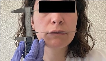
Figure 1. Measurement of the canthus–commissure distance.
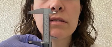
Figure 2. Measurement of the lower facial third at rest.
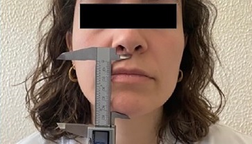
Figure 3. Measurement of the lower facial third in occlusion
Results
A total of 36 students were included in the study, with 12 participants from each academic year (3rd, 4th, and 5th year). The mean age was 22.4 years, and the gender distribution was 22% male and 78% female.
Among the three variables analyzed, the VDR showed the greatest variability (mean = 68.9 mm; SD = 7.3), whereas the CCD presented the least variability (mean = 63.5 mm; SD = 4.7) (Figure 4, Table 1).
The CCD showed a strong, positive, and statistically significant correlation with both the VDO (r = 0.701; p < 0.001) and mean value of the CCD lower than those of VDO (p < 0.001) and VDR (p < 0.001). These findings support a robust association among these facial dimensions, indicating that increases in one variable tends to correspond to increases in the others (Figures 5 and 6). Scatter plots with linear regression lines confirmed these associations, showing moderate to strong coefficients of determination (R² = 0.550 for VDO and R² = 0.501 for VDR). Despite these associations, the dispersion of data points and the presence of systematic differences between measurements suggest limited predictive accuracy. Comparative analysis between the CCD and both VDR and VDO revealed statistically significant differences, with the mean value of the CCD lower than those of VDO (p < 0.001) and VDR (p < 0.001).
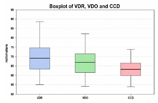
Figure 4. Box plot of facial dimension of the canthus–commissure (CCD), lower facial third at rest (VDR), lower facial third in occlusion (VDO).
Table 1. Descriptive statistics of the evaluated facial measurements.
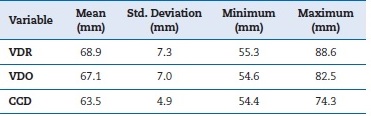
VDR – vertical dimension at rest; VDO – vertical dimension of occlusion; CCD – canthus–commissure distance
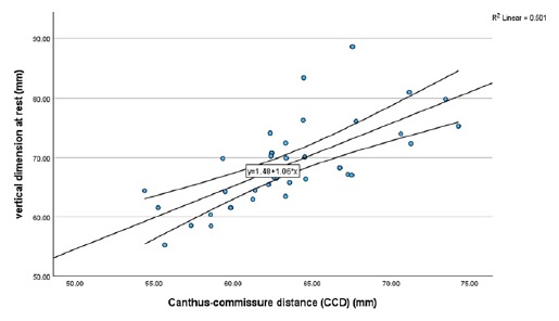
Figure 5. Scatter plot showing the relationship between the canthus–commissure distance and vertical dimension at rest, with linear regression line and 95% confidence interval.
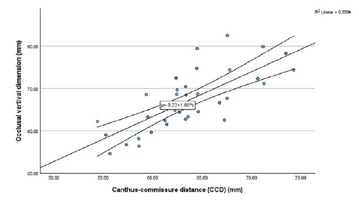
Figure 6. Scatter plot showing the relationship between the canthus–commissure distance and vertical dimension of occlusion, with linear regression line and 95% confidence interval.
Discussion
Determining the VDO is a crucial step in the rehabilitation of total edentulous patients, but it presents significant challenges due to the absence of fixed anatomical landmarks and high individual variability, which hinder the standardization of measurement methods.2, 4, 15 Errors in establishing the VDO— whether by overestimation or underestimation—may lead to temporomandibular joint dysfunction, denture instability or misfit, pain, compromised masticatory function, facial aesthetic alterations, and angular cheilitis.1 16, These complications negatively affect patients’ quality of life and may increase the need for subsequent adjustments or even result in prosthesis failure.
The Willis method is widely employed to determine the VDO because it is simple, non-invasive, and does not require sophisticated equipment. However, its definition remains ambiguous, as the lower third of the face is mobile and variable, introducing uncertainty regarding the precise dimension at which Willis’s proposed facial proportionality applies. In the present study, a strong and statistically significant positive correlation was observed between CCD and both VDR and VDO. Although this finding indicates that these variables change in a coordinated manner, it should not be interpreted as evidence of equivalence or interchangeability. The regression models further illustrate that, although a linear relationship exists, a substantial proportion of variability remains unexplained, reinforcing the limited reliability of using this method as a standalone predictor.
Several limitations should be acknowledged. First, the use of a convenience sample, justified by accessibility, data- collection efficiency, and participants’ familiarity with the study concepts, may introduce bias and limit generalizability. Moreover, the relatively young and homogeneous sample may not reflect the variability of typical prosthodontic populations, particularly older edentulous patients.
Additionally, an asymmetric sex distribution was observed in the sample, with a predominance of female participants, which likely reflects the growing feminization of dental education and the dental profession in Portugal; this also represents a limitation, as the sex imbalance may influence the interpretation and generalizability of the results. Because the study population consisted exclusively of young dentate individuals, extrapolation to edentulous or older populations should be made with caution, as soft-tissue
characteristics, neuromuscular control, and facial morphology may differ substantially.Another limitation was that measurement accuracy was influenced by soft-tissue variability—especially in individuals with convex facial profiles or facial hair—and by slight tissue compression associated with caliper use. These constraints are consistent with the broader limitations of direct facial measurements (e.g., Willis compass or calipers), which are inherently affected by soft-tissue compressibility and operator-dependent variability.
Additionally, VDR determination may be influenced by physiological factors such as posture, muscle relaxation, and breathing pattern, which should be considered when interpreting the results. Although cephalometry provides a more objective skeletal assessment, its static nature limits functional relevance. In contrast, digital approaches, including 2D/3D photogrammetry and facial scanning, enhance accuracy and reproducibility in the evaluation of facial thirds by reducing soft tissue distortion and enabling standardized landmark identification. While advanced 3D systems are not yet widely available in routine clinical practice, simplified methods using standardized smartphone photography may offer a pragmatic alternative, albeit with lower precision.
Clinically, these findings reinforce that the vertical dimension of occlusion determination should rely on a combination of anatomical, functional, and phonetic methods rather than any single measurement approach. Within this context, facial proportional methods should be considered as complementary tools rather than standalone determinants of vertical dimension.
These findings are consistent with previous reports of high variability in facial measurements, with alignment of facial thirds occurring in only 43.3% of participants.(7) Similarly, other studies have shown that alternative facial measurements, such as the distance from the pupil center to the lip junction, are highly variable in both dentate and edentulous patients. (14) Facial soft tissues are subject to individual variability influenced by age, sex, facial morphology, and muscle tone. These factors may partially explain why proportional equivalence is not consistently observed across individuals.
Conclusions
A statistically significant correlation exists between the canthus–commissure distance and lower facial thirds, both at rest and in occlusion. However, the canthus–commissure distance alone does not provide a sufficiently reliable parameter for accurately determining either the vertical dimension at rest or the vertical dimension of occlusion. Importantly, these findings should be interpreted with caution, as their generalizability to the broader population—particularly edentulous patients— is limited due to the use of a convenience sample composed exclusively of dentate individuals.
Within these constraints, the Willis method may still serve as an adjunctive clinical reference for estimating the vertical dimension of occlusion. However, its application should be interpreted with caution and should not be used as a standalone approach in prosthodontic rehabilitation. The presente study should be interpreted as an exploratory cross-sectional clinical investigation. Although the sample size was estimated from pilot continuous-variable data, the study was not designed to establish predictive models or definitive clinical agreement thresholds. Therefore, the findings should be interpreted with caution and confirmed in future studies involving larger and more heterogeneous samples, including edentulous populations, a more balanced sex distribution, and digital facial analysis methods to better define the clinical utility of facial proportional approaches.
References
1. Felton DA. Edentulism and comorbid factors. J Prosthodont. 2009;18(2):88-96.
2. Calamita M, Coachman C, Sesma N, Kois J. Occlusal vertical dimension: treatment planning decisions and management considerations. Int J Esthet Dent. 2019;14(2):166-181.
3. Sudhir N, Chittaranjan B, Kumar BA, Taruna M, Kumar MP, Reddy MR. Digital cephalometric tracings by PRO-CEPH V3 software for comparative analyses of vertical dimension in edentulous patients. J Clin Diagn Res. 2015;9(5):ZC01-5.
4. Espinosa-Valarezo JC, Irribarra-Mengarelli R, González-Bustamante H. Métodos de evaluación de la dimensión vertical oclusal. Rev Clin Periodoncia Implantol Rehabil Oral. 2018;11(2):116-120.
5. Gaspard M. Troubles de l’occlusion dentaire et SADAM. Collection du chirurgien-dentiste. Sèvres: Procodif Éditeur; 1985.
6. Willis FM. Esthetics of denture construction. J Am Dent Assoc. 1930;17:636-642.
7. Magalhães JB, Zavanelli AC, Zavanelli RA. Analysis of facial proportion and its relation with dental form, rest and occlusal vertical dimension between institutionalized students. Rev Odontol UNESP. 2011;40(5):215-221.
8. Fayz F, Eslami A. Determination of occlusal vertical dimension: a literature review. J Prosthet Dent. 1988;59(3):321-3.
9. Fayad MI, Moussa R, Ragheb NA, Ahmed Y, Sultan S, AbdulSalam MR, Ismail HA, Elboraey MO, AbdElaziz MH, Helal MA. Vertical dimension in prosthodontics theory and practice (Part II): a comprehensive review of vertical dimension determination in prosthodontics from classical methods to digital innovation. Cureus. 2025;17(10):e93987.
10. Russi S. Verificação experimental do método de Willis: contribuição ao estudo da dimensão vertical [doctoral thesis]. Araraquara: Faculdade de Farmácia e Odontologia de Araraquara; 1965.
11. Reis KR, Telles DM, Fried E, Kaizer OB, Bonfante G. Análise do método de Willis na determinação da dimensão vertical. Rev Bras Odontol. 2008;65(1):48-51.
12. Emam NM. Correlation between nose-to-chin distance and other measurements used to determine occlusal vertical dimension. Cureus. 2024;16(5):e60443.
13. Singh DK, Subhas S, Gupta A, Kesari M, Kumar A, Nayak L. Facial measurements: a guide for vertical dimension. J Family Med Prim Care. 2020;9(4):2056-60.
14. Nagpal A, Parkash H, Bhargava A, Chittaranjan B. Reliability of different facial measurements for determination of vertical dimension of occlusion in edentulous using accepted facial dimensions recorded from dentulous subjects. J Indian Prosthodont Soc. 2014;14(3):233-42.
15. Trentin LM, Reginato VF, Maroli A, Borges MTR, Spazzin AO, Bacchi A. Determinação da dimensão vertical de oclusão em prótese total: revisão de literatura e relato de caso clínico. J Oral Investig. 2016;5(1):50-60.
16. Zarb GA, Bolender CL, Hickey JC, Carlsson GE. Boucher’s Prosthodontic treatment for edentulous patients. 10th ed. St. Louis: Mosby Company; 1990.
Isabel Gomes
E-mail address: isabel.gomes@fmd.ulisboa.pt
CRediT authorship contribution statement
Isabel Gomes: Conceptualization, Investigation, Methodology, Project administration, Supervision, Validation, Visualization, Writing – review & editing. Cátia Faustino: Data curation, Formal analysis, Investigation, Visualization, Writing – original draft. Henrique Luís: Formal analysis, Methodology, Validation, Writing – review & editing. Luís Pires Lopes: Validation, Visualization, Writing – review & editing.
Ethical disclosures
Protection of human and animal subjects. The authors declare that the procedures followed were in accordance with the regulations of the relevant clinical research ethics committee and with those of the Code of Ethics of the World Medical Association (Declaration of Helsinki).
Confidentiality of data. The authors declare that no patient data appear in this article.
Right to privacy and informed consent. The authors have obtained the written informed consent of the patients or subjects mentioned in the article. The corresponding author is in possession of this document.
Conflict of interest
he authors declare that they have followed the protocols of their work center on the publication of patient data.
1646-2890/© 2026 Sociedade Portuguesa de Estomatologia e Medicina Dentária. Published by SPEMD.
This is an open access article under the CC BY-NC-ND license (http://creativecommons.org/licenses/by-nc-nd/4.0/).