
Revista Portuguesa de Estomatologia, Medicina Dentária e Cirurgia Maxilofacial
Rev Port Estomatol Med Dent Cir Maxilofac | 2017 | 58 (1) | 23-31
Original research
Aesthetic analysis of the face
Análise estética da face
a Department of Orthodontics, Faculty of Medicine, University of Coimbra, Coimbra, Portugal
b Department of Statistics, Faculty of Medicine, University of Coimbra, Coimbra, Portugal
Francisco do Vale - fvale@fmed.uc.pt, franciscofvale@gmail.com
Article Info
Rev Port Estomatol Med Dent Cir Maxilofac
Volume - 58
Issue - 1
Original research
Pages - 23-31
Go to Volume
Article History
Received on 10/05/2016
Accepted on 24/11/2016
Available Online on 31/03/2017
Keywords
Original research
Aesthetic analysis of the face
Análise estética da face
Francisco do Valea,*, Joana Queirogaa, Francisco Caramelob, Luísa Malóa, Pedro Leitãoa, João Maló-Abreua
aDepartment of Orthodontics, Faculty of Medicine, University of Coimbra, Coimbra, Portugal bDepartment of Statistics, Faculty of Medicine, University of Coimbra, Coimbra, Portugal
http://doi.org/10.24873/j.rpemd.2017.05.002
abstract
Objective: Using well-characterized sample of an european caucasian population, this study seeks to create lateral cephalometric information, based on the Natural Head Position within the assessed european population groups, to analyze sexual dimorphism and to study the comparison with similar work done in an american Caucasian group.
Methods:Fifty-five subjects (20 men and 35 women) were selected with the following criteria: caucasian descent, over 18 years old and presenting good facial aesthetics; Angle class I occlusion with no crowding and without temporomandibular disfunction; no previous history of orthodontic treatment or surgical interventions in the maxillofacial area. The following records were collected: (1) clinical data; (2) alginate impressions to obtain the study models; (3) four pictures in the Natural Head Position (NHP); (4) lateral cephalometric radiographs in centric occlusion and in the NHP. The american caucasian sample, 20 men and 26 women, were selected on the basis of the same criteria.
Results:The thickness of the skin tissues that cover the face is larger in men than in women and there are no major ethnic differences. The total face height is significantly higher in men than in women, and is higher in european women compared to North-america women. The midface is further back sagittally in men than in women and is further back in the ideal european population versus the ideal american population.
Conclusion: In a cephalometric analysis, we must consider the gender, age and etnic background of the patient in order to obtain correct clinical information.
Keywords: Cephalometry, Esthetics, Ethnic groups, Sex characteristics
Resumo
Objetivo: Este trabalho, recorrendo a amostras populacionais bem caracterizadas, procura criar informação cefalométrica lateral, baseada na Posição Natural da Cabeça e aferida a grupos de uma população caucasiana europeia. Também pretende analisar o dimorfismo sexual e estudar a comparação com trabalhos idênticos feitos numa população caucasiana norte-americana.
Métodos: Cinquenta e cinco indivíduos (20 homens e 35 mulheres) foram selecionados pelos seguintes critérios: caucasianos, maior de 18 anos, com boa estética facial; oclusão Classe I de Angle, sem apinhamento nem disfunção temporomandibular; sem história prévia de tratamento ortodôntico ou intervenções cirúrgicas na área maxilo-facial. Os seguintes registros foram recolhidos: (1) história clínica; (2) impressões em alginato para obter modelos de estudo; (3) quatro fotografias na posição natural da cabeça (PNC); (4) telerradiografias de perfil da face em oclusão cêntrica e em PNC. A amostra caucasiana americana, 20 homens e 26 mulheres, foi seleccionada com base nos mesmos critérios.
Resultados: A espessura dos tecidos cutâneos que recobrem a face é maior nos homens do que nas mulheres e não se verificam grandes diferenças étnicas. A altura total da face é significativamente maior nos homens do que nas mulheres, e é maior nas mulheres europeias em relação às mulheres norte-americanas. O andar médio da face está mais recuado sagitalmente nos homens do que nas mulheres e mais recuado na população europeia do que na população norte-americana.
Conclusão: Na interpretação da análise cefalométrica devem ser considerados o género, a idade e a etnia do indivíduo a estudar, para se obter uma correcta informação clínica.
Palavras-chave: Cefalometria, Estética, Grupos étnicos, Características sexuais
Introduction
Facial aesthetics are currently one of the main objectives of orthodontic treatment, together with a normal occlusion, healthy periodontal tissues and the stability of the treatment.
Orthodontics, beyond seeking a functional balance between bones and teeth within a cephalometric correction, also seeks to develop facial harmony as part of the treatment objectives.
Through cephalometrics, orthodontics acquires the capacity to detect abnormalities and measure the degree of dentoskeletal and soft tissue disharmony.
There are several published studies that fit cephalometric norms to various ethnic groups in an attempt to establish optimal standards of occlusion and facial aesthetics for these populations. Some authors1 - 3 emphasize the importance of soft tissue analysis in their publications. However, these types of studies on the Portuguese-Caucasian population are rare. Pereira4 compared a sample population of 12 year old Portuguese children with Caucasian groups of Norwegian origin.
This study found that the Portuguese children had longer and more convex faces than the Norwegian children. Leitão5 investigated and discussed the utility of natural head position–based cephalometric variables, evaluating the relationship between natural head position and craniofacial morphology.
Using a well-characterized sample of a Portuguese-Caucasian population, this study seeks to create lateral cephalometric information based on the Natural Head Position to analyze sexual dysmorphism and study the comparison with similar work done among other ethnic groups.
The objectives of this study are: to establish ideal cephalometric norms in lateral incidents of soft tissues that serve as a reference point for diagnostic, orthodontic or surgical-orthodontic treatment plan of an adult Portuguese-Caucasian population; to compare cephalometric norms obtained from men with those obtained from women and to compare cephalometric norms obtained from the studied Portuguese population with norms from North American caucasians obtained from the Arnett Soft Tissue Cephalometric
Analysis (STCA) at the Center for Corrective Jaw Surgery, Santa Barbara, CA.1 - 3
The following null hypotheses will be tested: there are no morphological differences between men and women; and there are no morphological differences between ethnic groups.
Materials and Methods
The study was approved by the Ethics Committee from the Faculty of Medicine of the University of Coimbra.
The ideal population sample was obtained from 578 young Portuguese-Caucasian adults of both sexes, students from Faculty of Medicine of the University of Coimbra and other volunteers.
The following criteria were used to select the patients for data collection: A normal class I molar and canine occlusion; the absence of facial marks, such as scarring, as well as mandibular functional deviations; to be of Portuguese-Caucasian origin; absence of previous orthodontic treatment or surgery in the maxillofacial area.
The sample was reduced to 59 people, and the following data was collected: clinical data; study models obtained from alginate impressions of the dental arches; photographs in numbers of four: two frontal and two profile of the Natural Head Position (NHP).
The following criteria were used to select the patients for the final ideal population sample:
– Presence of all permanent teeth; molar and canine class I,6 with overbite and overjet of 0.5 mm – 4 mm.
– Well-aligned teeth in the arches, without excess or missing space greater than 3mm per arch.
– The absence of: teeth with rotation or inclination more than 15º; ectopic eruption; supernumerary teeth; teeth in infra- or supra-occlusion; anterior or posterior crossbites; deviation of the dental midline relative to the facial midline greater than 0.5 mm; functional anterior or lateral deviations; forced bites, with deviations between the first tooth contact and maximum intercuspidation, greater than 1.5 mm;
– Without signs or symptoms of temporomandibular dysfunction;
– To have completed the period of bone growth. The chronological age must be over 18 years old;
– The presence of good overall facial harmony; clinical selection performed by two orthodontists and two postgraduate orthodontistry students from our school.
Fifty-five people (35 women and 20 men) constituted the final population. Their ages varied between 18 – 33 years with an average of 22.6 years of age. In addition to the aforementioned exams, this final population underwent lateral cephalometric radiography in the Natural Head Position.
The North-American sample1 - 3 is constituted of 46 young Caucasian adults (20 men and 26 women), selected according to a dental and facial aesthetic criteria: the entire population contained a Class I dental occlusion, without prior orthodontic treatment, and good facial aesthetics. The methods of execution and study of the cephalometric analysis followed by the american author are similar to the steps described below.
A number of instruments were used in the execution of the teleradiography in the Natural Head Position. These included: a Siemens Orthophos CD Panoramic Dental X-ray, Kodak 24x30 cm cassettes and Kodak 8DS1 18x24 cm films, and a Gevamatic 60 automatic developing machine. In addition, the cephalostat of the Panoramic Dental X-ray was adapted with a metal chain to project the radiographic film. On the wall, two meters in front of the cephalostat, an adjustable mirror (120x40 cm) was placed.
The lateral cephalometric radiographs were taken in centric occlusion. The centric relation was obtained through a standard manual manipulation and recorded on a wax bite which was used as an occlusal guide when taking the cephalometric radiography. In preparation for the cephalometric radiography, metal markers were used to better identify parts of the soft tissue structures of the midface, specifically: the Cheek Bone CB, Orbital Rim OR, Alar Base AB and Subpupil SP.
To obtain the NHP a test was first made to ensure the patient found a comfortable position of the head. This was followed by the positioning of the cephalostat. The final position was achieved by rearranging the feet, body and head, adjusting its symmetry with the metal chain.
The relaxation movements of the body and head were repeated during the test, paying attention to the reflection on the mirror. With the midface cutaneous points marked, as well as with the positioned condyles and the lips relaxed, a teleradiography was a conducted on the Natural Head Position.
The cephalometric analysis was performed with the direct digital method by Dolphin Imaging Software/32 (High Quality Digital Imaging Software for Orthodontics, Orthographic Surgery, Cosmetics and Medical Imaging), using the 8.0.6.12 version from the Dolphin Imaging Systems Inc, USA. The conversion of teleradiographies to digital images was done using the Astra 2400S HAL0 scanner with a transparency adapter (UMAX Technologies, Inc., Fremont, USA) and Photoshop 5.01® (Abode Systems Incorporated, San Jose, USA). To ensure exact measurements, the scan was obtained on a 100% scale with a resolution of 300 DPI without image enhancement filters. This method was chosen because it is the most popular choice by respected authors.7
Once scanned, the radiographic images were saved in JPEG format on a personal computer. The cephalometric values were subsequently exported to a spreadsheet.
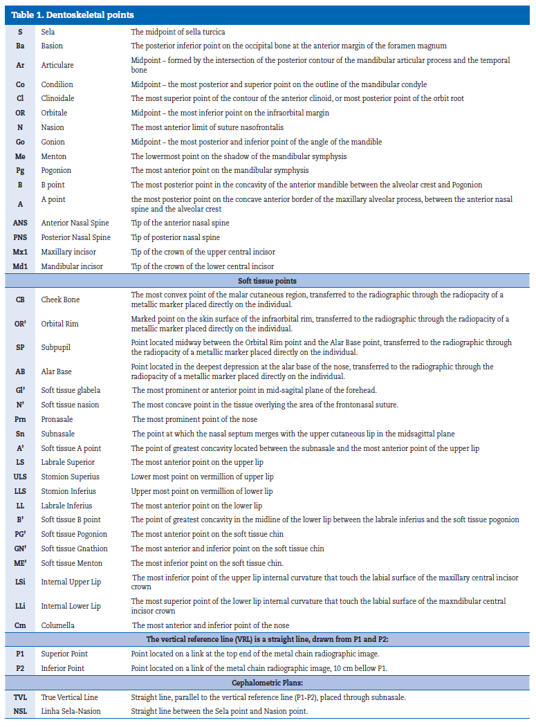
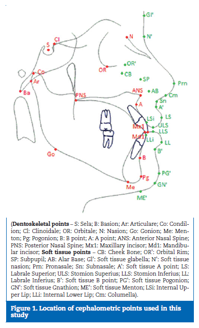
Table 1 describes and Figure 1 shows the location of all points used in this study.
The repetition of the cephalometric tracing was carried out by a direct digital method of 20 randomly selected lateral teleradiographies, three weeks following the initial cephalometric tracing by the same investigator. To detect any systematic errors, a t-test with a significance level of 5% was used for each pair of records8 (Table 2). The random error was studied using a formula proposed by Dahlberg.9 The Pearson correlation coefficient(r) was determined for each pair of records.
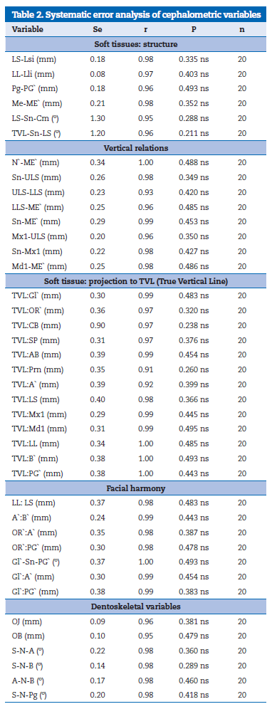
Table 3 and 4 present the results of variables that establish the cephalometric norms of the ideal Portuguese-Caucasian population, sexual dysmorphism and ethnic dysmorphism.
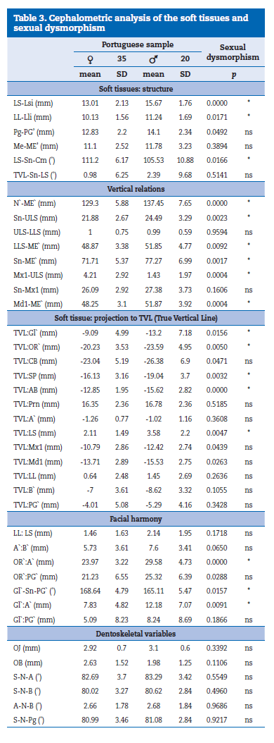
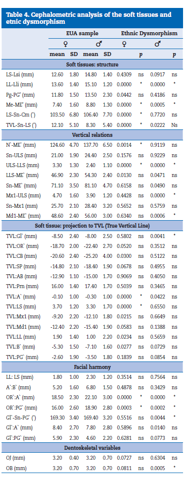
The thickness of the upper lip (LS-LSI) and lower lip (LL-Lli) is significantly different between both sexes and there is no difference between the two ethnic groups for the upper lip. Males presented a thicker upper lip and lower lip in comparison to females. The thickness of the Pogonion Cutaneous (Pg-PG`) is not significantly different between both sexes and neither between the two ethnic groups. The thickness of the Soft Tissue Menton (Me-ME`) shows no significant differences between both male and female, but is higher in the Portuguese sample in comparison to the North American sample. The naso-labial angle (LSSn- Cm) is significantly more open in females than in males in the Portuguese sample and also more open than in females of the North American sample. The upper lip angle (TVL-Sn-LS) is not significantly different between the two sexes in the Portuguese sample. However, it is significantly lower in Portuguese females in comparison to the North-american sample.
The total face height (N`-ME`) is significantly higher in males than in females, and higher among the females of the Portuguese sample versus North American females. The length of the upper lip (Sn-ULS) is significantly higher in males versus females in the Portuguese sample without any ethnic dysmorphism.
The length of the lower lip (LLS-ME`) and the height of the lower face (Sn-ME`) are significantly higher in males than females in the Portuguese sample. The exposure of the upper incisor (Mx1-ULS) is significantly lower in males than in females of the Portuguese sample and significantly lower in males of the Portuguese sample compared to males of the North American sample.
The Soft Tissue Glabella (TVL:Gl`) is significantly more posterior in males than females, and more posterior in the Portuguese male sample compared to the North-American male sample. The variables corresponding to the midface (TVL:OR`, TVL:SP, TVL:AB) are significantly more posterior in males than in females in the Portuguese sample and do not demonstrate ethnic dysmorphism. The anteroposterior position of the upper lip TVL:LS is more advanced in males (3.58 ±2.2 mm) than in females (2.11 ±1.49 mm), and in comparison to the North-american sample the lip recess in Portuguese women is significantly higher. The anteroposterior position of the lower lip TVL:LL is not statistically significant in both sexes of the Portuguese sample and do not demonstrate ethnic dysmorphism. The positioning of the upper incisors TVL:Mx1 and lower incisors TVL:Md1 is not statistically significantly different between both sexes in the Portuguese sample and neither between the two ethnic groups.
In the relationship between the lips LL:LS there are no significant differences either between the sexes nor between the two ethnic groups. Regarding the harmony between the soft-tissue landmarks that best represent the sagittal position of the jaw (A`:B`) it was not verified a statistically significant anteroposterior difference.
The overbite (OB) is significantly higher in the North American males than in the Portuguese males, while the overjet (OJ) has no significant difference between the two ethnic groups.
The A-N-B angle that describes the intermaxillary sagittal relationship is almost the same in both sexes, 2.66 ± 1.78º in females and 2.68 ± 1.84º in males.
Discussion
The differences in the structure and placement of the soft tissue profile is a result of many factors including heredity and environment. As the profile varies with the type of malocclusion, this study only included patients with a Class I occlusion, although there are aesthetically pleasing faces in patients with malocclusion. There may also be large skeletal variations in individuals with a class I molar and canine.
These considerations are some of the reasons why the study was conducted only with subjects possessing a normal occlusion associated with good facial aesthetics.
Males presented a thicker upper lip and lower lip in comparison to females. The highest growth of the lips is verified in girls between the ages of 10 and 14 and in boys between the ages of 8 and 1611. If the thickness of the upper lip LS-LSI is greater than 18mm, it does not follow the movement of the incisors during treatment. But if the thickness is less than 12mm, then, the lip is already tracking the incisive contraction. 12 , 13
The nasolabial angle decreases with age11 and can be modified either through the nose growth, either by sagittal changes of the lips during orthodontic or orthodontic-surgical treatment14.
The values found in this study (111 ± 6 females; males 105 ± 11) and the values of other authors5,15-20 demonstrate quite well the variability of this angle and consequently the interest in using different normative values in individuals of different populations.
The length of the upper lip (Sn-ULS) is significantly higher in males versus females in the Portuguese sample. The largest growth of the upper lip occurs in men between 10 and 16 years of age and in women between 10 and 14 years of age.21 The implication of a short lip in facial aesthetics is important because it causes a higher incisive exposition and a relative increase in the height of the lower face.
The labial measurements allow us to identify if the length of the soft tissue is normal or abnormal and, consequently, provides information about the length of structures of the dentoskeletal tissues. Farkas17 described an increased lip length of 0.77mm/year in men of 9 to 18 years of age and an increase of 0.46mm/year in girls of 8 to 16 years of age.
In this study, the interlabial gap (ULS-LLS), with relaxed lips, does not present significant differences between the two sexes which is consistent with other studies15,17-20. Regarding ethnic dysmorphism, the values found in the Portuguese sample were significantly lower than those presented in the North American sample. This measure depends on the length of the lips, skeletal vertical length, projection of the incisors and lip posture.21
The exposure of the upper incisor (Mx1-ULS) is significantly lower in males than in females of the Portuguese sample and significantly lower in males of Portuguese sample compared to males of the North American sample. Looking at the above data, it is noticeable that these differences relate to differences in the upper lip length. Subtelny22 in a study of 30 adolescents verified the existence of a constant vertical relationship between the incisal edge of the central incisor and the upper lip growth.
The nasal projection TVL:Prn is not significantly different between the sexes and between ethnic groups. Authors like Arnett and Bergman1, 2 and Lehman20 considered a big nose when projected to more than 20 mm and a small one when projected less than 14 mm. The values found in this investigation are within those ranges (16 ±2.36 mm).
The variables TVL:OR`, TVL:CB and TVL:SP are indicative of the sagittal position of the upper jaw and the TVL:AB variable demonstrates the anteroposterior position of the upper and lower jaw. The significant recess verified in males pertains to sexual differences of the facial structure, considering the greatest projection of cheekbones verified in females.
The facial contour angle Gl`-Sn-PG` relates the three portions of the face. For this variable, ethnic dysmorphism was not found in females, and it was verified a wider Gl`-Sn-PG` in females (168.64 ±4.79º) than in males (165.11 ±5.47º). These values are consistent with the values found by several authors.1,15, 16,18-20
The S-N-A angle oscillated between 83,29º in the male sample and between 82,69º in the female sample, thus, not presenting significant differences. These values are consistent with the values found by several authors.23-26
The two variables that indicate a sagittal jaw position, S-N-B e S-N-Pg, statistically were the same in both males and females; however, the male population has a slightly advanced position of the mandible. Other authors24,27 had significantly different values in both sexes. This may be justified because these authors did not include an aesthetic criteria as part of the selection process of their samples.
The results are to be understood only as reference values for a well-characterized population. Therefore, when comparing some of the variables with the results of other investigations it is natural to observe differences in the results that may not be entirely related to ethnicity. Some studies use different selection criteria for the population, some studies do not use the natural head position, and the exact way of measuring the same variable may be different from study to study.
Then, in a cephalometric analysis, we must consider gender, age and etnic background in order to obtain correct clinical information. The Natural Head Position, relaxed lips and the use of metallic markers to identify some parts of the soft tissue structures of mid-face are essential for proper cephalometric analysis when used as a diagnostic and treatment planning tool. The harmony between the constituent parts of the face is determined regardless of the True Vertical positioning.
Conclusions
1. In the Portuguese sample, males in comparation to females, presented: a higher total face height and a higher lower face height; more posterior position of soft tissue glabella and variables corresponding to the midface; thicker and longer upper and lower lips with a more advanced anteroposterior position of the upper lip; smaller naso-labial angle; and lower exposure of the upper incisors.
2. Females, in the Portuguese sample in comparison to the North American sample, presented: a higher naso-labial angle; higher total face height; lower upper lip angle; lower anteroposterior position of the upper lip. Males, in the Portuguese sample in comparison to the North American sample, presented: lower overbite; lower exposure of the upper incisor; more posterior soft tissue glabella. In the Portuguese sample in comparison to the North American sample, the thickness of the Soft Tissue Menton is higher in both genders.
3. If the cephalometric standards obtained in this study are accepted as ideal for the population, then, orthodontic treatment and/or orthodontic-surgical treatment based on dental-skeletal standards is not sufficient to obtain good facial aesthetics and could even lead to undesirable clinical outcomes.
References
1. Arnett GW, Bergman RT. Facial Keys to orthodontic diagnosis and treatment planning-Part I. Am J Orthod 1993;103:299-312.
2. Arnett GW, Bergman RT. Facial Keys to orthodontic diagnosis and treatment planning-Part II. Am J Orthod 1993;103:395-411.
3. Arnett GW, Jelic SJ, Kim J, Cummings DR, Beress A, Worley M, Chung B, Bergman R. Soft tissue cephalometric analysis: Diagnosis and treatment planning of dentofacial deformity. Am J Orthod Dentofac Orthop 1999;116:239-53.
4. Pereira R. Face Morphology of 12 year old children in Portugal (MS Thesis). Bergen: University of Bergen, Faculty of Dentistry, 1993.
5. Leitão P, Nanda RS. Relationship of natural head position to craniofacial morphology. Am J Orthod Dentofac Orthop 2000;17:406-17.
6. Angle EH. Classification of malocclusion. Dent Cosmos 1899;41:248-64,350-7.
7. Ongkosuwito EM, Katsaros C, Van`T Hof MA, Bodegon JC, Kuijpers-Jagtman AM. The reproducibility of Cephalometric measurements: a comparison of analogue and digital methods. Europ J Orthod 2002;24:655-65.
8. Houston WJB. The analisys of errors in orthodontic measurements. Am J Orthod 1983;83:382-9.
9. Dahlberg G. Statistical methods for medical and biological students. New York: Interscience Publications, 1940.
10. Benjamini, Y. & Yekutieli, D. 2001 The control of the false discovery rate in multiple testing under dependency. The Annals of Statistics;294:1165-88.
11. Mamandras HF. Linear changes of maxilary and mandibular lips. Am J Orthod 1988;94:405-10.
12. Holdaway R A. A soft-tissue cephalometric analysis and its use in orthodontic treatment planning Part I 1983. Am J Ortho;84:1-28.
13. Holdaway R A. A soft-tissue cephalometric analysis and its use in orthodontic treatment planning Part II 1984. Am J Orthod;85:279-93.
14. Farkas LG, Kolar JC. Anthropometrics and art in the aesthetic of women`s faces. Clin Plast Surg 1987;14:599-615.
15. Burstone CJ. Integumental contour and extension patterns. Angle Orthod 1959;29:93-104.
16. Burstone CJ. Lip posture and its significance in treatment planning. Am J Orthod 1967;53:262-84.
17. Farkas LG.Anthropometry of the head and face in medicine. New York: Elsevier North Holland Inc. 1981.
18. Legan HL, Burstone CJ. Soft-tissue cephalometric analysis for orthognathic surgery. Oral Surg 1980;38:744-51.
19. Powell N, Humphreys B. Proportions of the esthetic face. New York: Thieme-Stratton, 1984.
20. Lehman JÁ. Soft tissue manifestations of the jaws: diagnosisand treatment. Clin Plast Surg 1987;14:767-83.
21. Mamandras HF. Linear changes of maxilary and mandibular lips. Am J Orthod 1988;94:405-10.
22. Subtelny JD. A longitudinal study of soft tissue facial structures and their profil characteristics defined in relation to undrelying skeletel structures. Am J Orthod Dentofac Orthop 1959;45:381-507.
23. Björk A. The Face in Profile – an anthropological x-ray investigation on Swedish children and conscripts. Sven Tandlak Tidskr 1947;40(Supp 5B):1-180.
24. McNamara JA, Ellis E. Cephalometric analysis of untreated adults with ideal facial and occlusal relationships. J Adult Orthod Orthog surg 1988;3:221-31.
25. Cerci V, Martins J, Olivcira M. Cephalometric standards for white Brazilians: Int J Adult Orthod Orthognath Surg 1993;8:287-22.
26. Leitão P. Contribuição para o estudo das características craniofaciais da população Portuguesa (Tese Doutoramento). Lisboa: Universidade de Lisboa,Faculdade de Medicina Dentária. 1997.
27. Segner D. Floating norms as a means to describe skeletal patterns. Eur J Orthod 1989;11:214-20.
Francisco do Vale
E-mail address: fvale@fmed.uc.pt , franciscofvale@gmail.com
Ethical disclosures
Protection of human and animal subjects. The authors declare that the procedures followed were in accordance with the regulations of the relevant clinical research ethics committee and with those of the Code of Ethics of the World Medical Association (Declaration of Helsinki).
Confidentiality of data. The authors declare that no patient data appear in this article.
Right to privacy and informed consent. The authors declare that no patient data appear in this article.
Conflict of interest
The authors have no conflicts of interest to declare.
Article history:
Received 10 May 2016
Accepted 24 November 2016
Available online 30 March 2017