
Revista Portuguesa de Estomatologia, Medicina Dentária e Cirurgia Maxilofacial
SPEMD | 2019 | 60 (3) | 96-103
Original research
Bonding performance of a universal adhesive: effect of hydrophobic resin coating and long-term water storage
Desempenho de um sistema adesivo universal: efeito da aplicação de uma camada de resina hidrófoba e do envelhecimento em água
a Instituto de Dentisteria Operatória, Área de Medicina Dentária, Faculdade de Medicina, Universidade de Coimbra
b Laboratório de Bioestatística e Informática Médica, Faculdade de Medicina, Universidade de Coimbra
Alexandra Vinagre - avinagre@fmed.uc.pt
Article Info
Rev Port Estomatol Med Dent Cir Maxilofac
Volume - 60
Issue - 3
Original research
Pages - 96-103
Go to Volume
Article History
Received on 03/08/2019
Accepted on 15/10/2019
Available Online on 04/11/2019
Keywords
Original research
Bonding performance of a universal adhesive: effect of hydrophobic resin coating and long-term water storage
Desempenho de um sistema adesivo universal: efeito da aplicação de uma camada de resina hidrofoba e do envelhecimento em água
Alexandra Vinagrea,*, Ana Ralhoa, Nuno Ramosa, Ana Messiasb, João Carlos Ramosa
a Instituto de Dentisteria Operatória, Área de Medicina Dentária, Faculdade de Medicina, Universidade de Coimbra
b Laboratório de Bioestatística e Informática Médica, Faculdade de Medicina, Universidade de Coimbra
http://doi.org/10.24873/j.rpemd.2019.11.463
Abstract
Objectives: To evaluate the immediate and water-aged microtensile bond strength of a universal adhesive to dentin, using the self-etch approach with and without an additional hydrophobic resin layer.
Methods:Flat dentin surfaces were prepared from twelve non-carious human molars and were randomly divided into two groups. The universal adhesive system Scotchbond™ Universal was used in self-etch mode (SBU) with and without the application of an extra hydrophobic resin layer of Adper™ Scotchbond™ Multi-Purpose Plus (SBU+HL). After composite buildups, samples were stored in water (37oC/7 days) and then sectioned into microspecimens (1.00±0.2 mm2). Half of the microspecimens were immediately subjected to microtensile bond strength testing (0.5 mm/min) while the other half was stored in water, according to the ISO/TS 11405:2015, for 4 years before testing. Data were analyzed with Kruskal Wallis and all-pairwise comparisons with Bonferroni corrections (p<0.05).
Results:The following microtensile bond strengths were registered (mean in MPa±SD): immediate SBU, 52.5 ±13.56; 4-year SBU, 26.0±6.50; immediate SBU+HL, 59.3±12.39; 4-year SBU+HL, 32.5±5.72. No statistically significant differences were detected between adhesive strategies either immediately or in the aged period (p>0.05). A significant decrease in bond strength was verified between the immediate and the 4-year evaluation for both groups (p<0.05).
Conclusions:Water storage induced an approximately 50% reduction in dentin bond strength, regardless of the adhesive strategy employed. The incorporation of an extra hydrophobic layer over the universal adhesive did not improve dentin bond strength significantly either immediately or after long-term water storage.
Keywords: Aging, Dentin, Microtensile bond strength, Universal adhesives
Resumo
Objetivos: Avaliar as forcas de adesão por microtração a dentina de um adesivo universal aplicado no modo autocondicionante, com ou sem uma camada de resina hidrofóbica.
Métodos: Foram preparadas superfícies de dentina em 12 molares, divididos aleatoriamente em dois grupos. O sistema adesivo Scotchbond™ Universal foi aplicado no modo autocondicionante (SBU) ou com a adição de uma camada extra de resina hidrofóbica Adper™ Scotchbond™ Multi-Purpose Plus (SBU+HL). Após a restauração em resina composta, os dentes foram armazenados em água (37oC/7 dias) e seccionados em bastonetes (1,00±0,2 mm2). Metade dos especimes foi sujeita ao teste de resistência adesiva a microtração (0,5 mm/min) enquanto que os restantes foram armazenados em água por 4 anos segundo as normas ISO/TS 11405:2015. Para análise estatística foi utilizado o teste de Kruskal Wallis e as comparações em pares foram realizadas com as correcções de Bonferroni (p<0,05).
Resultados: Os resultados da resistência adesiva foram os seguintes (media em MPa±SD): SBU_imediato 52,5±13,56; SBU_4 anos 26,0±6,50; SBU+HL_imediato 59,3±12,39; SBU+HL_4 anos 32,5±5,72. Não foram encontradas diferenças estatisticamente significativas entre as estratégias adesivas quer no período imediato ou após o envelhecimento (p>0,05). Foi verificado um decréscimo significativo da resistência adesiva do período imediato para os 4 anos para ambos os grupos (p<0,05).
Conclusões: O armazenamento em água induziu uma redução de cerca de 50% das forcas de adesão a dentina, independentemente da estratégia utilizada. A aplicação da camada de resina hidrofóbica não melhorou significativamente as forcas de adesão quer na avaliação imediata ou após 4 anos de envelhecimento em água.
Palavras-chave: Envelhecimento, Dentina, Microtração, Adesivos universais
Introduction
The limited durability of restorations results mainly from the exposure of adhesive interfaces to the oral environment, where they are permanently subjected to mechanical, chemical and/or thermal stimuli.1 The main challenge in adhesive dentistry is to bond effectively to substrates of different natures.
Bonding to enamel is reliable and durable. In contrast, bonding to dentin is still challenging due to its variable nature and heterogeneous structure.2, 3 Current adhesive technologies tend to produce simplified materials with reduced clinical application times and decreased technique sensitivity.4
Contemporary dental adhesive systems can be classified according to their application techniques as etch-and-rinse and self-etch adhesive systems. Self-etch adhesive systems contain acidic monomers that simultaneously etch and infiltrate the dental substrate, discarding the highly sensitive step of acid etching.5, 6 The composition and concentration of the acidic resin monomers establish differences in the acidity and aggressiveness of these systems, determining distinct bonding interfacial ultra-morphologies.3,5,7 Their aggressiveness depends on the pH of the solution, and self-etch systems can be categorized according to their pH into ultra-mild (pH > 2.5), mild (pH ≈ 2), intermediately strong (pH between 1 and 2) and strong (pH ≤ 1).3
Universal or multi-mode adhesive systems were introduced more recently and can be applied either with the etchand-rinse or the self-etch mode. This multi-approach capability allows the clinician to apply the adhesive with the so-called selective enamel etching technique, which combines the advantages of an etch-and-rinse technique on enamel with a simplified self-etch approach on dentine.4, 5, 8, 9 The development of universal adhesives followed the all-in-one concept of the existing one-step self-etch adhesives, thus requiring water in their formulation to ionize hydrophilic acidic monomers.10 Most often, carboxylate and/or phosphate groups are their primary functional monomer. Due to its reported high performance, 10-methacryloyloxydecyl monomer (10-MDP) has been incorporated in a wide range of adhesives in this class. The 10-MDP molecule is an amphiphilic functional monomer with a long carbonyl chain backbone between the functional and the polymerizable groups in the monomer structure, which renders it fairly hydrophobic.11 Moreover, when applied on dentin surfaces, it forms self-assembled nanolayers of hydrolytically stable calcium salts, which explain its high bond stability.12, 13 Recent data evidenced that the bond-strength stability of these adhesives to dentin depends largely on their pH, emphasizing that mild universal adhesives are less dependent on the adhesive strategy used and are materials with better stability after aging. Nevertheless, dentin bond strength mediated by ultra-mild and intermediately strong universal adhesives decrease significantly with aging, particularly when an etch-and-rinse strategy is employed.9
The instability of the dentin adhesive interfaces of simplified one-step self-etch and universal adhesives has been attributed to the presence of permeable hybrid layers that allow the circulation of water throughout the interface after polymerization.
This permeability favors water sorption by polymers and progressive hydrolytic and enzymatic degradation of the unprotected collagen by matrix metalloproteinases (MMPs).14, 16
To overcome these problems, modified adhesive formulations and/or application techniques have been evaluated.16, 20 The use of an extra hydrophobic resin coat after universal adhesive application aims to increase the thickness of the adhesive layer and homogenize it, as well as reduce the fluid flow across the adhesive interface and decelerate bond degradation.20
The aim of this study was to evaluate the immediate (7 days) and water-aged (4 years) dentin microtensile bond strength of a universal adhesive employed using the self-etch approach with and without an additional hydrophobic resin layer. The hypotheses tested were the following: (1) the application of an extra hydrophobic resin layer over a light-cured universal adhesive does not improve immediate or aged dentin bond strength, and (2) the bonding efficiency of both adhesive strategies does not decrease after 4 years of water aging.
Material and methods
Twelve caries-free, intact human third molars were collected following ethical approval (Ethical Committee of the Faculty of Medicine of Coimbra, Portugal; CE-001/2013). The teeth were stored in a 0.5% chloramine solution at 4ºC for up to 6 months after extraction, cleaned of all debris and partially included in a self-curing acrylic resin block (Vertex, Vertex-Dental, Zeist, Netherlands). The occlusal surfaces were cut with a diamond saw perpendicularly to the long axis of the tooth (Accutom 5, Struers, Ballerup, Denmark), under water-cooling, thereby exposing a flat mid-coronal dentin surface. All exposed surfaces were wet-ground with a sequence of 200–,400- and 600-grit silicon-carbide sandpaper in a circular motion for 60 seconds each to standardize smear layer preparation,21 and were carefully observed under a stereomicroscope (Nikon® SMZ 1500, Tokyo, Japan) to confirm the absence of residual enamel and other defects.
The teeth were randomly assigned to two groups (n=6). In group I (SBU), dentin adhesive procedures were performed according to the manufacturer’s directions with a multi-mode adhesive system in the self-etch mode: ScotchbondTM Universal Adhesive (3M ESPE, St. Paul, MN, USA). In group II (SBU+HL), bonding procedures started similarly, but an additional hydrophobic resin layer (AdperTM ScotchbondTM Multi-Purpose Adhesive, 3MEspe, St. Paul, MN, USA) was applied after (Table 1).
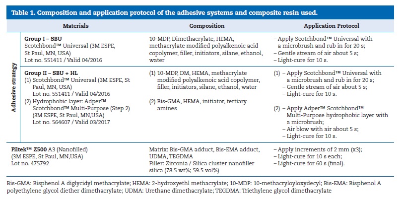
After adhesive procedures, resin composite buildups were prepared using a Filtek™ Z500 A3 (3M ESPE, St. Paul, MN, USA) microhybrid composite in 2-mm increments to a height of 6 mm (Table 1). Each layer was light-cured for 10 seconds, followed by a final polymerization of 60 seconds using a LED light-curing unit (Bluephase 20i®, Ivoclar Vivadent, Schann, Lichtenstein) at a power density of 1080 mW/cm2 measured using a digital radiometer (Bluephase® Meter II, Ivoclar Vivadent, Schann, Lichtenstein). The teeth were stored for 7 days in distilled water at 37°C (Heraeus BK 6160, Kelvitron® Kp, Wehrheim, Germany).
Afterward, the specimens were sectioned longitudinally across the bonded interface in mesiodistal and buccal-lingual directions with a low-speed saw (Accutom 5, Struers, Ballerup, Denmark) at 300 rpm, under refrigeration, to obtain composite- adhesive-dentin sticks with a cross-sectional area of approximately 1.00 ± 0.2 mm2, as measured using a digital caliper (Digimatic Caliper, Mitutoyo; Tokyo; Japan). After the first cut in the x-axis direction, the free residual space between the slices was filled with the light-body silicone Aquasil Ultra XLV (Dentsply, DeTrey, Konstanz, Germany). For each tooth, the tops of adjacent sticks were identified with two colors. Half of the peripheral and central sticks were used to measure microtensile bond strength (μTBS) after 7-day storage and the others to determine mily:CaeciliaLTStd-Roman; mso-bidi-font-family:CaeciliaLTStd-Roman;color:black'>μTBS after 4-year storage in water at 37oC under the same protocol. During these 4 years, the medium was replaced every seven days to avoid contamination, following the ISO/TS 11405:2015 recommendations.22 All sticks were checked on an optical microscope (M300, Leica, Switzerland) at 40x magnification to exclude faulty specimens.
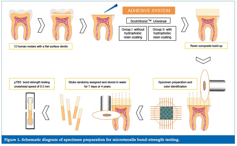
Each stick was attached to a microtensile sample holder with cyanoacrylate adhesive (CE10FlexR, Ce Chem Limited, Derbyshire, UK) and then fixed on the microtensile device (Od04-Plus; Odeme Dental Research, Luzerna, Brazil). Specimens were fractured in tensile mode using a universal testing machine (Model AG-I, Shimadzu Corporation, Kyoto, Japan) at a crosshead speed of 0.5 mm/min. The maximum load was recorded in Newtons, and microtensile bond strength was calculated in MPa according to the following equation: μTBS = F/A, where F is the load at fracture (N) and A is the bonded area (mm2). Figure 1 shows a schematic diagram of tooth preparation, restoration, specimen sectioning and bond-strength testing.
The failure mode was analyzed under an optical microscope (Leica CLS 150 MR, Switzerland) at 40x magnification.
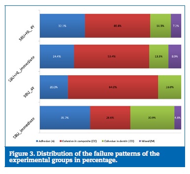
The fracture pattern was classified as follows: adhesive, if the failure occurred entirely within the adhesive interface; cohesive, if it occurred completely in the composite resin (cohesive in the resin) or the dentin (cohesive in the dentin); and mixed, when both adhesive and cohesive failure occurred.
Statistical analysis was performed with IBM SPSS 23.0R software (SPSS; Chicago, IL, USA). The normality of data was assessed with the Kolmogorov-Smirnov test and visual inspection of the histograms. Homoscedasticity was assessed with Levene’s test. Since these two assumptions were not verified, the Kruskal-Wallis test was run to determine the effect of the adhesion strategy and aging on bond strength, followed by all-pairwise comparisons with Bonferroni corrections. The chisquare test was used to compare the distribution of failure modes between groups. For all analyses, the significance level was set at α=0.05.
Results
The overall mean μTBS, standard deviations, number of specimens (n) and multiple comparison statistical analyses of all experimental groups are detailed in Table 2 and represented in Figure 2.
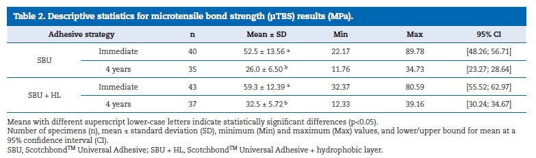
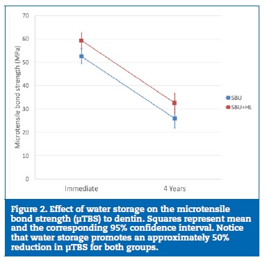
Data assessed with the Kolmogorov-Smirnov test failed to prove the normality of bond-strength values. The Kruskal-Wallis test determined that the distribution of bond-strength values across groups was not similar (p<0.001). All pair-wise comparisons showed no statistically significant differences in μTBS between adhesive strategies in both periods (p=0.504 for immediate and p=0.408 for 4-year aging). Both adhesive strategies showed a significant decrease in bond strength between the immediate and the 4-year evaluation (p<0.001). Both evaluation periods showed superior microtensile bond strength when the universal adhesive was coated with an extra hydrophobic resin layer, although the difference was not statistically significant. Figure 3 shows the distribution of the failure patterns of the experimental groups. No statistically significant differences were detected in the distribution of the failure mode, as evidenced by the chi-square test. The most common failure mode was cohesive for all adhesive strategies employed.
Discussion
Universal or multi-mode adhesives represent the latest generation of adhesive systems and have been developed under the ‘‘all-in-one’’ concept of the already existing one-step selfetch adhesives. Concerning most universal adhesives, in vitro studies report consistently that a similar immediate dentin microtensile bond strength can be achieved using either the etch-and-rinse or the self-etch adhesive strategy while exhibiting a relatively high performance.4, 23 Nevertheless, when adhesive interfaces were subjected to aging processes, several studies concluded that the self-etch approach resulted in more stable long-term bond characteristics.6, 9, 24 One meta-analysis of in vitro studies that evaluated nanoleakage related to universal adhesives reported divergent results and highlighted that etching modes influenced significantly and dissimilarly the nanoleakage of universal adhesives. Namely, the etch-and-rinse technique significantly reduced the nanoleakage of the Peak Universal and G-Bond Plus adhesives, whereas the self-etch mode reduced the nanoleakage of All-Bond Universal. In comparison, the etching technique did not significantly modify the nanoleakage pattern of either Prime&Bond Elect or Scotchbond Universal, which presented the lowest nanoleakage.25
The mean immediate dentin μTBS obtained in the present study with the application of ScotchbondTM Universal, an ultra-mild adhesive (pH=2.7), using the self-etch mode is in agreement with previously reported data at the upper threshold, in which bond strengths ranged from 32.3 to 59.9 MPa. In previous studies, when the same adhesive system was applied in the etch-and-rinse mode, μTBS was within a similar range of values (32.3 to 55.7 MPa).5, 17, 20, 26 - 29 In this study, the group with the application of an additional hydrophobic resin layer showed better bond-strength values both immediately and after 4 years of water storage. However, for both periods, no statistically significant differences were detected between adhesive strategies. Therefore, the first null hypothesis is rejected.
Several studies indicated a beneficial effect on dentin bond strength with the application of an extra hydrophobic adhesive layer over ScotchbondTM Universal or other adhesive systems.17, 19, 20, 30 - 32 A recent paper highlighted that the overall effect of using an extra bonding layer on the immediate and aged dentin bonding strength of the more recently introduced universal adhesives was dependent on the specific adhesive and its application mode.33 The application of an extra hydrophobic resin layer has been suggested to improve the adhesion of universal adhesives to dentin since it adds unsolvated hydrophobic monomers to the adhesive interface, which decreases the relative concentration of retained solvents and unreacted monomers in the adhesive layer. Furthermore, the increased thickness of this layer and the improved monomer conversion may also contribute to an increase in the dentin microtensile bond strength of the adhesive interface, due to the formation of a more densely packed hybrid layer, which renders it less permeable to water and less prone to degradation effects over time.17, 20, 31, 34 Ermis et al.20 measured dentin microtensile bond strength of universal adhesives immediately and after 6 months of water aging, with and without an additional hydrophobic layer applied over a separately light-cured or non-lightcured universal adhesive. They found that if the universal adhesive was used as a primer and was not light-cured before the application of the extra hydrophobic resin layer, no significant increase in bond strength was recorded, thus proving that this thicker adhesive layer impaired solvent volatilization, negatively affecting bond strength. Therefore, the bond durability of universal systems would only benefit from the application of an extra hydrophobic layer if the universal adhesive were separately light-cured. However, this polymerization strategy was employed in the present study and caused no significant improvement on bond strength.Several studies focusing on long-term bonding efficiency showed that adhesives suffer from bond degradation upon artificial and accelerated aging.6, 19, 26, 33, 35 Those findings are in line with the results of the present study, meaning the second null hypothesis is rejected. After 4 years of water storage, a significant drop in bond-strength values was observed both in the SBU (26.0±6.50 MPa) and the SBU+HL groups (32.5±5.72 MPa). Most studies evaluate long-term bonding performance of universal adhesives after at most one year of water storage, while this study focuses on a 4-year aging period. Considering that specimens were directly exposed to water and accelerated hydrolysis was expected, relatively acceptable bondstrength values were still found for both adhesive strategies in this experimental worst-case scenario. ScotchbondTM Universal contains 10-MDP as its acidic functional monomer and offers a low demineralization capacity.
Its interaction with dentin is either micromechanical, through shallow but adequate dentin hybridization,23, 27 or chemical, through the establishment of a strong ionic bond between 10-MDP and calcium atoms within hydroxyapatite crystals, forming a highly organized layer of stable and low-soluble calcium-phosphate salts.12, 13 Nevertheless, recent findings demonstrate that the hybrid layer stability of resin-dentin bonds in 10-MDP-based adhesives has been wrongly attributed to the presence of these nanolayered structures with insoluble MDP-calcium salts.36 Tian et al.36 evidenced that, after 1 year of water aging, although nano-layering features were identified in the dentin interface where 10-MDP and primer were applied, bond strength decreased significantly, and suggested that binding between calcium salts and the dentin surface had become weaker. In resin composites, inorganic fillers are silanized with methacryloxy silanes to allow their bond to the methacrylate resin matrix. In the case of MDP-calcium salts, the inward-facing of the methacrylate groups of two 10-MDP molecules may drastically reduce the number of freely available methacryloxy functionalities for coupling to the resin matrix, and this can explain the deterioration of the chemical interaction.36 Furthermore, the presence of polyalkenoic acid copolymer in the composition of SBU may compete with the 10-MDP by binding to the calcium of the hydroxyapatite.37
Apart from impairing the bonding of this functional monomer to dentine, the polyalkenoic acid copolymer may also have prevented monomer approximation during polymerization due to its high molecular weight, by diminishing the degree of conversion and the hybridization quality. 28
On the other hand, ScotchbondTM Universal contains the monofunctional monomer hydroxyethyl methacrylate (HEMA), a low molecular weight water-soluble monomer that behaves as a solvent by improving the miscibility and solubility of monomers with different polarity features. Besides, HEMA can also stabilize the collagen fibril network, improving the dentinal permeability, wettability and monomer diffusion.38 However, the presence of HEMA further increases the hydrophilic nature of self-etch adhesives as it retains water and diminishes its polymerization efficiency. In fact, HEMA promotes water uptake from the underlying dentin through osmosis, thus inducing the presence of water droplets on the surface of the adhesive layer that behaves as a semi-permeable membrane.39, 40 This high hydrophilicity weakens the polymer mechanical strength, making it more prone to degradation.41
Furthermore, HEMA has been documented to reduce nano-layering of 10-MDP at adhesive interfaces.42
The combination of all the above-mentioned factors may explain the loss of bonding efficiency and integrity of adhesive dentin interfaces over time, after water aging. Therefore, the contribution of chemical bonding to the overall bonding durability requires further investigation.
The determination of the most favorable application mode for universal adhesives remains debatable. Randomized clinical trials evidenced that using an etch-and-rinse or a self-etch approach to bond resin composite restorations in non-carious cervical lesions with universal adhesives did not significantly influence their clinical performance regarding marginal adaptation, marginal discoloration, post-operative sensitivity and secondary caries. Nevertheless, improved survival rates are linked to the use of the etch-and-rinse or the selective enamel etching procedure.43, 44 A recent clinical trial indicated that Scotchbond Universal applied with the self-etch mode induced a relatively high level of marginal discoloration.45
Further in vitro and, mainly, in vivo experiments should be carried out to understand better the degradation mechanisms of universal adhesives on dentin using differential and complementar aging methods, and to evaluate the long-term clinical performance of this newest category of adhesive systems.
Conclusions
Within the limitations of the present study, the results indicated that water storage induced an approximately 50% reduction in dentin bond strength, regardless of the adhesive strategy employed. The incorporation of an extra hydrophobic layer over a universal adhesive system did not improve dentin bond strength significantly either immediately or after long-term water storage.
References
1. De Munck J, Van Landuyt K, Peumans M, Poitevin A, Lambrechts P, Braem M, et al. A critical review of the durability of adhesion to tooth tissue: methods and results. J Dent Res. 2005;84:118-32.
2. Breschi L, Mazzoni A, Ruggeri A, Cadenaro M, Di Lenarda R, De Stefano Dorigo E. Dental adhesion review: aging and stability of the bonded interface. Dent Mater. 2008;24:90-101.
3. Van Meerbeek B, Yoshihara K, Yoshida Y, Mine A, De Munck J, Van Landuyt KL. State of the art of self-etch adhesives. Dental Mater. 2011;27:17-28.
4. Rosa WL, Piva E, Silva AF. Bond strength of universal adhesives: A systematic review and meta-analysis. J Dent. 2015;43:765-76.
5. Wagner A, Wendler M, Petschelt A, Belli R, Lohbauer U. Bonding performance of universal adhesives in different etching modes. J Dent.
6. Marchesi G, Frassetto A, Mazzoni A, Apolonio F, Diolosa M, Cadenaro M, et al. Adhesive performance of a multi-mode adhesive system: 1-year in vitro study. J Dent. 2014;42:603-12.
7. Perdigao J, Lopes MM, Gomes G. In vitro bonding performance of self-etch adhesives: II–ultramorphological evaluation. Oper Dent. 2008;33:534-49.
8. Perdigao J, Kose C, Mena-Serrano AP, De Paula EA, Tay LY, Reis A, et al. A new universal simplified adhesive: 18-month clinical evaluation. Oper Dent. 2014;39:113-27.
9. Cuevas-Suarez CE, da Rosa WLO, Lund RG, da Silva AF, Piva E. Bonding Performance of Universal Adhesives: An Updated Systematic Review and Meta-Analysis. J Adhes Dent. 2019;21:7-26.
10. Dieng-Sarr F, Sharrock P, Dabsie F, Gregoire G. Modifications of the organic and mineral fractions of dental tissues following conditioning by self-etching adhesives. J Dent. 2011;39:141-7.
11. Van Landuyt KL, Snauwaert J, De Munck J, Peumans M, Yoshida Y, Poitevin A, et al. Systematic review of the chemical composition of contemporary dental adhesives. Biomaterials. 2007;28:3757-85.
12. Yoshihara K, Yoshida Y, Nagaoka N, Fukegawa D, Hayakawa S, Mine A, et al. Nano-controlled molecular interaction at adhesive interfaces for hard tissue reconstruction. Acta Biomater. 2010;6:3573-82.
13. Yoshihara K, Yoshida Y, Nagaoka N, Fukegawa D, Hayakawa S, Mine A, et al. Nano-controlled molecular interaction at adhesive interfaces for hard tissue reconstruction. Acta Biomater. 2010;6:3573-82.
14. Tekce N, Tuncer S, Demirci M, Balci S. Do matrix metalloproteinase inhibitors improve the bond durability of universal dental adhesives? Scanning. 2016;38:535-44.
15. Serkies KB, Garcha R, Tam LE, De Souza GM, Finer Y. Matrix metalloproteinase inhibitor modulates esterase-catalyzed degradation of resin-dentin interfaces. Dent Mater. 2016;32:1513-23.
16. Comba A, Maravic T, Valente L, Girlando M, Cunha SR, Checchi V, et al. Effect of benzalkonium chloride on dentin bond strength and endogenous enzymatic activity. J Dent. 2019;85:25-32.
17. Munoz MA, Sezinando A, Luque-Martinez I, Szesz AL, Reis A, Loguercio AD, et al. Influence of a hydrophobic resin coating on the bonding efficacy of three universal adhesives. J Dent. 2014;42:595-602.
18. Luque-Martinez IV, Perdigao J, Munoz MA, Sezinando A, Reis A, Loguercio AD. Effects of solvent evaporation time on immediate adhesive properties of universal adhesives to dentin. Dent Mater. 2014;30:1126-35.
19. Sezinando A, Luque-Martinez I, Munoz MA, Reis A, Loguercio AD, Perdigao J. Influence of a hydrophobic resin coating on the immediate and 6-month dentin bonding of three universal adhesives. Dent Mater. 2015;31:e236-46.
20. Ermis RB, Ugurlu M, Ahmed MH, Van Meerbeek B. Universal Adhesives Benefit from an Extra Hydrophobic Adhesive Layer When Light Cured Beforehand. J Adhes Dent. 2019;21:179-88.
21. Ramos JC, Soares A, Torres S, Costa AL, Messias A, Vinagre A. Adhesive interface and microtensile bond strength evaluation of four adhesive systems to primary dentin. Ver Port Estomatol Med Dent Cir Maxilofac. 2016;57:65-73.
22. ISO/TS 11405:2015 – Dentistry–testing of adhesion to tooth structure. 2015.
23. Elkaffas AA, Hamama HHH, Mahmoud SH. Do universal adhesives promote bonding to dentin? A systematic review and meta-analysis. Restor Dent Endod. 2018;43:e29.
24. Lezaja Zebic M, Dzeletovic B, Miletic V. Microtensile bond strength of universal adhesives to flat versus Class I cavity dentin with pulpal pressure simulation. J Esthet Restor Dent. 2018;30:240-8.
25. Kaczor K, Gerula-Szymanska A, Smektala T, Safranow K, Lewusz K, Nowicka A. Effects of different etching modes on the nanoleakage of universal adhesives: A systematic review and meta-analysis. J Esthet Restor Dent. 2018;30:287-98.
26. Chen C, Niu LN, Xie H, Zhang ZY, Zhou LQ, Jiao K, Chen JH, Pashley DH, Tay FR. Bonding of universal adhesives to dentine–Old wine in new bottles? J Dent. 2015;43:525-36.
27. Perdigao J, Sezinando A, Monteiro PC. Laboratory bonding ability of a multi-purpose dentin adhesive. Am J Dent. 2012;25:153-8.
28. Munoz MA, Luque I, Hass V, Reis A, Loguercio AD, Bombarda NH. Immediate bonding properties of universal adhesives to dentine. J Dent. 2013;41:404-11.
29. Munoz MA, Luque-Martinez I, Malaquias P, Hass V, Reis A, Campanha NH, et al. In vitro longevity of bonding properties of universal adhesives to dentin. Oper Dent. 2015;40:282-92.
30. Chasqueira AF, Arantes-Oliveira S, Portugal J. Effect of changes to the manufacturer application techniques on the shear bond strength of simplified dental adhesives. J Appl Biomater Funct Mater. 2013;11:e117-21.
31. Perdigao J, Munoz MA, Sezinando A, Luque-Martinez IV, Staichak R, Reis A, et al. Immediate adhesive properties to dentin and enamel of a universal adhesive associated with a hydrophobic resin coat. Oper Dent. 2014;39:489-99.
32. Dal’ling MF LA, Bauer J, Carneiro KGK. Effect of an hydrophobic layer on a universal adhesive. RGO, Rev Gauch Odontol. 2018;66:339-44.
33. Ahmed MH, De Munck J, Van Landuyt K, Peumans M, Yoshihara K, Van Meerbeek B. Do Universal Adhesives Benefit from an Extra Bonding Layer? J Adhes Dent. 2019;21:117-32.
34. Reis A, Albuquerque M, Pegoraro M, Mattei G, Bauer JR, Grande RH, et al. Can the durability of one-step self-etch adhesives be improved by double application or by an extra layer of hydrophobic resin? J Dent. 2008;36:309-15.
35. Vermelho PM, Reis AF, Ambrosano GMB, Giannini M. Adhesion of multimode adhesives to enamel and dentin after one year of water storage. Clin Oral Invest. 2017;21:1707-15.
36. Tian F, Zhou L, Zhang Z, Niu L, Zhang L, Chen C, et al. Paucity of Nanolayering in Resin-Dentin Interfaces of MDP-based Adhesives. J Dent Res. 2016;95:380-7.
37. Yoshida Y, Yoshihara K, Nagaoka N, Hayakawa S, Torii Y, Ogawa T, et al. Self-assembled Nano-layering at the Adhesive interface. J Dent Res. 2012;91:376-81.
38. Moszner N, Salz U, Zimmermann J. Chemical aspects of self-etching enamel-dentin adhesives: a systematic review. Dent Mater. 2005;21:895-910.
39. Van Landuyt KL, Snauwaert J, De Munck J, Coutinho E, Poitevin A, Yoshida Y, et al. Origin of interfacial droplets with one-step adhesives. J Dent Res. 2007;86:739-44.
40. Gregoire G, Guignes P, Nasr K. Effects of dentine moisture on the permeability of total-etch and one-step self-etch adhesives. J Dent. 2009;37:691-9.
41. Cadenaro M, Antoniolli F, Sauro S, Tay FR, Di Lenarda R, Prati C, et al. Degree of conversion and permeability of dental adhesives. Eur J Oral Sci. 2005;113:525-30.
42. Yoshida Y, Yoshihara K, Hayakawa S, Nagaoka N, Okihara T, Matsumoto T, et al. HEMA inhibits interfacial nano-layering of the functional monomer MDP. J Dent Res. 2012;91:1060-5.
43. Arbildo H, Lamas-Lara C, Cruzado-Oliva F, Rosa-Prado C, Gomez-Fuertes A, Vasquez-Rodrigo H. Comparison of the clinical effect of the adhesive strategies of universal adhesives in the treatment of non-carious cervical lesions. Systematic review and meta-analysis. J Oral Res. 2018;7:210-22.
44. Nagarkar S, Theis-Mahon N, Perdigao J. Universal dental adhesives: Current status, laboratory testing, and clinical performance. J Biomed Mater Res B Appl Biomater. 2019;107:2121-31.
45. Ruschel VC, Shibata S, Stolf SC, Chung Y, Baratieri LN, Heymann HO, et al. Eighteen-month Clinical Study of Universal Adhesives in Noncarious Cervical Lesions. Oper Dent. 2018;43:241-9.
Alexandra Vinagre
E-mail address: avinagre@fmed.uc.pt
Ethical disclosures
Protection of human and animal subjects. The authors declare that no experiments were performed on humans or animals for this study.
Confidentiality of data. The authors declare that no patient data appear in this article.
Right to privacy and informed consent. The authors declare that no patient data appear in this article.
Conflict of interest
The authors have no conflicts of interest to declare.
Article history:
Received 3 August 2019
Accepted 15 October 2019
Available online 4 November 2019